A LITTLE BACKGROUND
The terms osteogenesis and
ossification are often used synonymously to indicate the
process of bone formation. Parts of
the skeleton form during the first few weeks after conception.
By the end of the eighth week after conception, the skeletal
pattern is formed in cartilage and connective tissue membranes
and ossification begins.
Bone development continues throughout adulthood. Even after
adult stature is attained, bone development continues for
repair of fractures and for remodeling to meet changing
lifestyles. Osteoblasts, osteocytes
and osteoclasts are the three cell types involved in the
development, growth and remodeling of bones. Osteoblasts
are bone-forming cells, osteocytes are mature bone cells
and osteoclasts break down and reabsorb bone.
There are two types of ossification: intramembranous and
endochondral.
Intramembranous ossification
involves the replacement of sheet-like connective tissue
membranes with bony tissue. Bones formed in this manner
are called intramembranous bones. They include certain flat
bones of the skull and some of the irregular bones. The
future bones are first formed as connective tissue membranes.
Osteoblasts migrate to the membranes and deposit bony matrix
around themselves. When the osteoblasts are surrounded by
matrix they are called osteocytes.
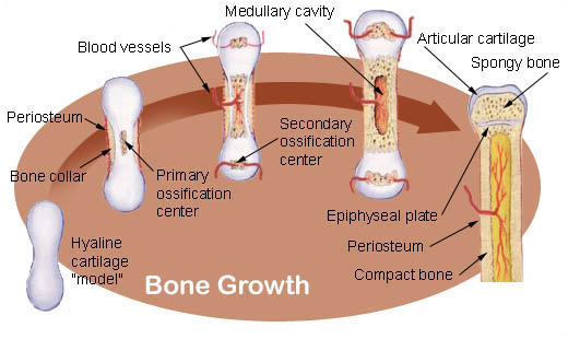
Endochondral ossification involves
the replacement of hyaline cartilage with bony tissue. Most
of the bones of the skeleton are formed in this manner.
These bones are called endochondral bones. In this process,
the future bones are first formed as hyaline cartilage models.
During the third month after conception, the perichondrium
that surrounds the hyaline cartilage "models"
becomes infiltrated with blood vessels and osteoblasts and
changes into a periosteum. The osteoblasts form a collar
of compact bone around the diaphysis. At the same time,
the cartilage in the center of the diaphysis begins to disintegrate.
Osteoblasts penetrate the disintegrating cartilage and replace
it with spongy bone. This forms a primary ossification center.
Ossification continues from this center toward the ends
of the bones. After spongy bone is formed in the diaphysis,
osteoclasts break down the newly formed bone to open up
the medullary cavity.
The cartilage in the epiphyses continues to grow so the
developing bone increases in length. Later, usually after
birth, secondary ossification centers form in the epiphyses.
Ossification in the epiphyses is similar to that in the
diaphysis except that the spongy bone is retained instead
of being broken down to form a medullary cavity. When secondary
ossification is complete, the hyaline cartilage is totally
replaced by bone except in two areas. A region of hyaline
cartilage remains over the surface of the epiphysis as the
articular cartilage and another area of cartilage remains
between the epiphysis and diaphysis. This is the epiphyseal
plate or growth region.
Bone Growth. Bones grow in
length at the epiphyseal plate by a process that is similar
to endochondral ossification. The cartilage in the region
of the epiphyseal plate next to the epiphysis continues
to grow by mitosis. The chondrocytes, in the region next
to the diaphysis, age and degenerate. Osteoblasts move in
and ossify the matrix to form bone. This process continues
throughout childhood and the adolescent years until the
cartilage growth slows and finally stops. When cartilage
growth ceases, usually in the early twenties, the epiphyseal
plate completely ossifies so that only a thin epiphyseal
line remains and the bones can no longer grow in length.
Bone growth is under the influence of growth hormone from
the anterior pituitary gland and sex hormones from the ovaries
and testes.
Ref:
http://training.seer.cancer.gov/module_anatomy/unit3_3_bone_growth.html
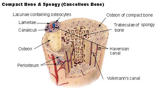
There are two types of bone
tissue: compact and spongy. The names
imply that the two types of differ in density, or how tightly
the tissue is packed together. There are three types of cells
that contribute to bone homeostasis. Osteoblasts are bone-forming
cell, osteoclasts resorb or break down bone, and osteocytes
are mature bone cells. An equilibrium between osteoblasts
and osteoclasts maintains bone tissue.
Ref: http://training.seer.cancer.gov/module_anatomy/unit3_2_bone_tissue.html
Some
definitions:
Hematopoiesis,
the formation of blood cells, mostly takes place in the
red marrow of the bones.
Alkaline
phosphatase:
An enzyme produced in liver and bone.
Going away from the center of ossification is the zone of
calcification where the cartilage is calcifying, a zone
of hypertrophy where the cartilage
is elongating, a zone of proliferation where new
cartilage cells are being produced, and finally an area
of hyaline cartilage. The area consisting of the zone of
ossification, calcification, hypertrophy, and proliferation
is termed the epiphyseal plate. http://www.auburn.edu/academic/classes/zy/0301/Topic7/Topic7.html
See
Gray's Anatomy - The Interior of the Skull:
http://www.bartleby.com/107/47.html
|
