Wallerian
degeneration
Reference
Synonyms:
Orthograde degeneration, secondary degeneration.
Associated
persons:
Augustus Volney WallerDescription:
Degeneration
of the distal segment of a peripheral nerve fibre (axon)
that has been severed from its nutritive centres (cell
body), without local inflammation. The myelin sheath
also degenerates, from a distal injury to the same axon,
and is transformed into a chain of stainable lipoid
droplets (Marchi's stain). This discovery made it possible
to trace the course of fibres through the nervous system
and demonstrated the importance of the nucleus in the
regeneration of nerve fibres. The neurilemma does not
degenerate but form a tube that directs the growth of
the regenerating axon.
Bibliography:
•
A. V. Waller:
Experiments on the section of the glossopharyngeal and
hypoglossal nerves of the frog, and observations on
the alterations produced thereby in the structure of
their primitive fibres.
London, Edinburgh and Dublin Philosophical Magazine
and Journal of Science, 1850.
Philosophical Transactions of the Royal Society of London,
1850, 140: 423-429.
Edinburgh Medical and Surgical Journal, 1851, 76: 369.
Recherches
sur la système nerveux.
Comptes rendus de l’Académie des sciences,
Paris, 1851,33: 370-374 and 606-611.
Observations
sur les effets de la section des racines spinales et
du nerf pneumogastrique au dessus de son ganglion inférieur
chez les mammifères.
Comptes rendus hebdomadaires des séances de l’Académie
des Sciences, Paris, 1852, 34: 582-587.
Sur
la reproduction des nerfs et sur la structure et les
fonctions des ganglions spinaux.
Archiv für Anatomie, Physiologie und wissenschaftliche
Medicin, Leipzig, 1852:392.
Experience
sur les sections des nerfs et les alterations.
Comptes-rendus de la Société de biologie,
Paris, 1857, 2 (3): 6.
|
Research
Project:
Organophosphate Insecticide Damage to the Mature
and Developing Nervous Systems: in Vitro Systems for Detection
and Remediation
Principal
Investigator: E.
Tiffany-Castiglioni
Address:
Veterinary Anatomy
Texas A&M University
College Station, Texas 77843
Start
Date:
04/2001
End Date: 04/2006
CRIS RPAs: 723 314 711
Excerpts:
... At ten times the concentration of mipafox that causes
a 50% inhibition of NTE (5x10⁻⁵ M/day) mipafox was found
to significantly decrease neurite
length in differentiated cells while paraoxon and
OPH-hydrolyzed paraoxon at the same concentration did
not.
...
While some organophosphorus (OP) compounds including paraoxon
produce acute toxicity through acetylcholinesterase inhibition,
others such as mipafox produce OP-induced delayed neurotoxicity
(OPIDN), which is characterized anatomically by Wallerian-type
"dying back" neuropathy in the axon and myelin.
...
Protein expression of NF200 was
shown to be a new biomarker by which the neurotoxic
effects of mipafox and paraoxon on SY5Y cells were distinguishable
at the molecular level.
...
The current study shows that organophosphorus compounds
produce not only antiesterase activity but also modifications
in protein. Evidence presented suggests that mipafox
caused shortening of neurites in differentiated SY5Y cells
by a degeneration process, whereas paraoxon inhibited
neurite growth in the cells.
Ref:
http://www.tard.state.tx.us/index.php?mode=Listing&rl_id=639
|
...
In a study of alkyl phosphate poisoning, Pasi and Leuzinger
came to the conclusion that delayed lesions only occur,
if at all, after severe cerebral anoxia [176]. As
regards anatomical changes in the brain (demyelination),
these delayed lesions correspond to those caused by
peripheral neuropathy in acute and chronic ortho-tricresyl
phosphate poisoning and
are confined to fluorine- containing alkyl phosphates
- for example, mipafox, DFP,
sarin and soman.
A
synoptic evaluation of 536 civilian cases of alkyl phosphate
poisoning made by the above-mentioned authors led them
to the conclusion that acute poisoning by civilian alkyl
phosphates did not result in delayed lesions. It should
be noted, however, that their period of observation
of two to three years was inadequate for investigations
of delayed lesions beside the scale of Spiegelberg and
others [page 40].
Ref: Delayed Toxic Effects of
Chemical Warfare Agents. A SIPRI (Stockholm international
Peace Research Institute) Monograph. 1975. ISBN 91-85114-29-4.
http://projects.sipri.se/cbw/research/cw-delayed.pdf
|
From Toxline at Toxnet
Journal
of Applied Toxicology, Vol. 13, No. 2, pages 143-145,
17 references, 1993
Delayed
Neurotoxic Effect of Sarin
in Mice after Repeated Inhalation Exposure
Husain
K, Vijayaraghavan R, Pant SC, Raza SK, Pandey KS
Abstract:
The ability of sarin (107448) to induce delayed neurotoxicity
was examined in mice. Female Swiss-albino-mice were
exposed to 5mg/m3 sarin vapor 20 minutes/day for 10
days. Other mice were injected subcutaneously with 2.5mg/kg
mipafox (371868) daily for 10 days. Mice were
observed for clinical signs of toxicity for 14 days
starting after the first sarin or mipafox exposure.
They were killed on day 14. Brain and spinal cord tissues,
and blood platelets were assayed for neurotoxic-esterase
(NTE) activity. Spinal cord sections were prepared and
examined for histopathological changes. Mice
exposed to sarin developed muscular weakness in the
limbs and ataxia on day 14. Mipafox exposed mice developed
severe ataxia. Both sarin
and mipafox inhibited brain, spinal cord, and platelet
NTE activity. Sarin was less potent than mipafox.
Sarin and mipafox induced spinal
cord axonal degeneration. The
degree of degeneration was greater in mipafox treated
mice. Sarin also caused focal axonal degeneration in
the lateral branches of the spinal cord. The authors
conclude that sarin seems capable of inducing delayed
neurotoxicity in mice following repeat inhalation exposure.
|
From Toxline at Toxnet
Human
and Experimental Toxicology, Vol. 16, No. 2, pages 67-71,
11 references, 1997
The
Effects of Multiple Low Doses of Organophosphates on
Target Enzymes in Brain and Diaphragm
in the Mouse
Williams
FM, Charlton C, de Blaquiere GE, Mutch E, Kelly SS,
Blain PG
Abstract:
The effects of multiple low doses of ecothiopate (513100),
paraoxon (311455), and mipafox
(371868) on organophosphate target enzymes in the brain
and diaphragm were studied in mice. Male albino-mice
were injected subcutaneously once with 0
or 110 micromoles per kilogram (micromol/kg) mipafox,
0.5micromol/kg ecothiopate, or 1.5micromol/kg paraoxon
or with 0 or 44micromol/kg mipafox,
0.2micromol/kg ecothiopate, or 0.6micromol/kg paraoxon
daily for 5 days or 27.5micromol/kg mipafox daily for
8 day s. The mice were killed 3 hours (hr) after the
single dose or 3 or 24hr after each of the multiple
doses and the brain and diaphragms were removed. The
brains were assayed for acetylcholinesterase (AChE)
and neuropathy-target-esterase (NTE) activity. The diaphragms
were analyzed for AChE activity. The
single doses of mipafox, ecothiopate, and paraoxon inhibited
diaphragm AChE activity to about the same extent, around
67 to 68%. Mipafox and paraoxon inhibited brain AChE
activity by 55 and 73%, respectively. Ecothiopate did
not affect brain AChE activity.
Only mipafox inhibited brain NTE activity, by 66%. Brain
AChE activity was progressively inhibited by 17 to 46%
by multiple dosing with 27.5micromol/kg mipafox, by
23 to 49% by multiple injection with 44micromol/kg mipafox,
and by 20 to 55% by multiple dosing with paraoxon.
Ecothiopate caused a small increase in brain AChE activity.
Diaphragm AChE activity was similarly
inhibited by mipafox and paraoxon. Ecothiopate
also caused a progressive inhibition of diaphragm AChE
activity. Brain and diaphragm AChE activity showed some
recovery between the daily doses. The extent of inter
dose recovery was greater in the case of paraoxon and
ecothiopate than with mipafox. Mipafox
also produced a progressive inhibition of brain NTE
activity, the cumulative inhibitory effect, 74 and 76%,
being similar after the two dosing protocols. The
authors conclude that exposure to multiple low doses
of mipafox, ecothiopate, and paraoxon produces additive
inhibition of AChE activity. These results have
implications for humans as humans are generally exposed
to low levels of organophosphates for extended periods
of time.
|
From Toxline at Toxnet
In
Vitro Toxicology. Journal of Molecular and Cellular
Toxicology, Vol. 5, No. 3, pages 127-136, 28 references,
1992
Cytotoxic
Effects of Organophosphorus Esters and Other Neurotoxic
Chemicals on Cultured Cells
Nostrandt
AC, Rowles TK, Ehrich M
Abstract:
The in-vitro cytotoxicity of organophosphates and other
neurotoxic chemicals in a neuronal cell line was examined.
Differentiated SY-5Y cells, a human neuroblastoma cell
line, were incubated with 0 to 10(-3) molar (M) mipafox
(371868), paraoxon (311455), aldicarb (116063), beta,beta-iminodipropionitrile
(111944) (IDPN), carbachol (51832), carbaryl (63252),
or phenyl-saligenin-phosphate (4081236) (PSP) for 24
hours. Other cells were incubated with the nonneurotoxicants
atropine or verapamil for comparison. Effects on viability
were determined using the trypan-blue dye exclusion
test. SY-5Y cells were incubated with mipafox, paraoxon,
aldicarb, IDPN, or carbachol for up to 14 days. In some
experiments, atropine was added to the cultures. The
cultures were assayed for acetylcholinesterase (AChE)
activity after 10 minutes. The cells were analyzed for
intracellular calcium (Ca+2) content after 3, 10, 24,
and 48 hours. The cultures were examined for histomorphological
changes periodically for up to 14 days. A parallel experiment
using chicken brain homogenates was performed and compared
with the effects on Sy-5Y AChE activity. Only
mipafox, carbachol, carbaryl, and PSP significantly
decreased cellular viability, with carbaryl and
PSP being the most potent. SY-5Y cells exposed to paraoxon,
aldicarb, or carbachol became rounded, more refractile,
and eventually detached from the culture plate after
3 days incubation. Mipafox induced
swelling and blebbing on the neurites after 3 days.
The neurites were significantly
shorter and thinner on day ten compared to the control
cultures. Significant numbers
of rounded and detached cells were seen on day 14. No
histopathological changes were seen in IDPN treated
cells until day ten, at which point they showed changes
similar to those induced by mipafox. Carbachol, aldicarb,
paraoxon, IDPN, and mipafox inhibited
AChE activity to a greater extent in SY-5Y cells than
in the chicken brain homogenates, with mipafox,
paraoxon, and aldicarb being the most potent.
Mipafox, paraoxon, aldicarb, IDPN, and carbachol induced
transient increases in Ca+2 content at 4 hours. Peak
Ca+2 concentrations occurred at 10 hours, except in
the case of paraoxon. Ca+2 concentrations in paraoxon
treated cells decreased sharply after 4 hours. Atropine
attenuated the increases in Ca+2 concentration induced
by the compounds. The authors
conclude that SY-5Y cells can be used to assess the
cytotoxic effects of neurotoxic chemicals, especially
esterase inhibitors.
|
The
reports are available from The National Technology Information
Service (NTIS) .
Order by: phone at 1-800-553-NTIS (U.S. customers); (703-)605-6000
(other countries); fax at (703)-605-6900; and email at orders@ntis.gov.
NTIS is located at 5285 Port Royal Road, Springfield, VA,
22161, USA. |
| Details |
Abstract |
Order
Number: NTIS/01930011
(4 pages)
2004
-
Atomic Crystal Structure of an Organophosphorus
Acid Anhydrolase.
Authors:
Quiocho FA, Nickitenko A
Baylor
Coll. of Medicine, Houston, TX.
Keywords:
Crystal structure
*Organophosphates
Three dimensional
X ray spectra
Chemical warfare agents
Crystallography
Enzyme inhibitors
Anhydrolases
X ray crystallography
Opaa(Organophosphorus acid anhydrolase) |
Final
progress rept. 5 Dec 2000-4 Dec 2003.
The major aim is to determine the three-dimensional atomic
structure of an organophosphorus acid anhydrolase (OPAA)
by x ray crystallography. This structure is a prerequisite
for remodeling the active site, in collaboration with scientists
at ECBC and Geo-Centers, Inc. in order to enhance catalytic
activity towards fluorinated U-type and other extremely
toxic chemical warfare (CW) nerve agents. A decontamination
system based on the remodeled OPAA not only provides rapid
removal of CW agents, but is also enviromnentally safe and
noncorrosive in nature. After overcoming the major difficulty
in crystallizing OPAA, we have now determined by MAD and
SAD techniques and refined the crystal structure of OPAA
to 2.5 A resolution. The structure
analysis of OPAA with the bound enzyme inhibitor MIPAFOX
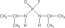
(N,N'-di-isopropyl phosphorodiamidic fluoride) is in progress. |
Order
Number: NTIS/AD-A290
426/6 (17 pages)
1994
- Genetic and Biochemical Manipulation
of a Broad-Spectrum Organophosphate Degrading System.
Authors:
Wild JR
Texas
A and M Research Foundation, College Station.
Keywords:
Genetics
Organophosphates
Pesticides
Pseudomonas |
Recent
studies on the plasmid-borne organophosphorus-degrading
gene of Pseudomonas diminuta and its enzyme have sought
to define both the genetic organization and the protein
chemistry involved in this system. The bacterial gene encodes
a single, unique enzyme, a phosphotriesterase (organophosphorus
anhydrase), which is capable of hydrolyzing a wide spectrum
of organophosphorus neurotoxins ranging from insecticides
such a parathion, orthene, coumaphos and diazinon to mammalian
neurotoxins such as diisopropylfluorophosphate (DFP), sarin,
soman and mipafox. The organophosphorus degrading genes
(opd) from two different plasmids in the soil bacteria P.
diminuta and Flavobacterium have been sequenced andtheir
structural organizations are being characterized. The cloned
geneshave been expressed in a number of biological systems
from bacteria to insect tissue culture, and the enzyme has
been purified and characterized from several different sources.
The catalytic reaction hasbeen determined to involve [abstract
truncated] |
Order
Number:
NTIS/PB94-137155 (7 pages)
1993
-
Differential Cytotoxic Sensitivity
in Mouse and Human Cell Lines Exposed to Organophosphate
Insecticides.
Authors:
Veronesi B, Ehrich M
Health
Effects Research Lab., Research Triangle Park, NC. Neurotoxicology
Div.
Virginia-Maryland
Regional Coll. of Veterinary Medicine, Blacksburg, VA.
Keywords:
Organophosphate insecticides
Toxicity
|
Neuroblastoma
cell lines were used to examine the differential interspecies
response (i.e., species selectivity) to organophosphates
(OPs). Baseline activities of the major target esterases,
i.e., cholinesterase, carboxylesterase, and neurotoxic esterase,
were assayed in mouse and several human neural candidate
cell lines. These activities were found to be variable within
individual cell lines and among the various tested cell
lines. Cytotoxicity data using the neutral red fluorometric
assay were collected on both human (SH-SY5Y) and mouse (NB41A3)
neuroblastoma clones exposed to a variety of OP insecticides.
IC50 data indicated that the tested mouse cell line was
consistently more sensitive than the human cell line to
equimolar doses of various OP compounds (e.g., mipafox,
parathion, paraoxon, DFP, leptophos oxon, fenthion, and
fenitrothion). These data suggest that interspecies-selectivity
in response to OP-related cytotoxicity is influenced by
intercellular differences in metabolism and basel [abstract
truncated] |
Order
Number: NTIS/PB95-126462
(1 8 pages)
1993
- Short-Term Clinical and Neuropathologic
Effects of Cholinesterase Inhibitors in Rats.
Authors:
Ehrich M, Shell L, Rozum M, Jortner BS
Virginia-Maryland
Regional Coll. of Veterinary Medicine, Blacksburg, VA.
Virginia
Polytechnic Inst. and State Univ., Blacksburg. Dept. of
Statistics.
Supporting Agency:
Health Effects Research Lab., Research Triangle Park,
NC.
Keywords:
Cholinesterase inhibitors
Nervous system
Pathology
Organophosphate insecticides
|
Adult
male Long Evans rats were given a single administration
of 3 dosage levels of the organophosphorus compounds tri-ortho-tolyl
phosphate (TOTP), diisopropyl fluorophosphate (DFP), phenyl
saligenin phosphate (PSP), mipafox, malathion, and dichlorvos
or the carbamate carbaryl. Acetylcholinesterase and neurotoxic
esterase activities were inhibited in a dose-dependent manner,
with the highest dosages of all these compounds inhibiting
activities of these enzymes in brain by at least 37% and
64%, respectively, at 4 and 48 hours after administration.
Rats given the high doses of TOTP (1000 mg/kg), DFP (3 mg/kg),
malathion (2000 mg/kg), and carbaryl (160 mg/kg) weighed
significantly less than control rats 14 days after administration.
A functional observational battery (FOB) was used to screen
for neurotoxic effects 1, 2, and 3 weeks after exposure.
All 7 test compounds were capable of causing changes in
parameters indicative of behavioral and central nervous
system excitability. In addition, dose-r [abstract truncated] |
Order
Number:
NTIS/PB93-229508 (10 pages)
1993
- Using Neuroblastoma Cell Lines
to Examine Organophosphate Neurotoxicity.
Authors:
Veronesi B, Ehrich M
Health
Effects Research Lab., Research Triangle Park, NC.
Virginia-Maryland
Regional Coll. of Veterinary Medicine, Blacksburg, VA.
Keywords:
Toxicity
Nervous system
Neuroblastoma
Organophosphate insecticides |
The
paper describes the initial characterization of neuroblastoma
cell lines to address several aspects of organophosphate
neurotoxicity. Several commercially available human and
mouse cell lines (i.e., SY5Y, IMR-32, SK-N-MC, NB41A3) were
evaluated for their target esterase activities (i.e., cholinesterase,
neurotoxic esterase, carboxylesterase), and of these cells,
a human (SY5Y) and mouse (NB41A3) neuroblastoma cell line
clone were used to establish an IC50 cytoxicity profile
for a variety of organophosphates insecticides (e.g., parathion,
paraoxon, diisopropylphosphorofluoridate and mipafox). The
human neuroblastoma cell line clone (SY5Y) was further used
to distinguish between neuropathy-causing OPs and cholinesterase
inhibitors. These initial data support the use of neuroblastoma
cell lines as effective test models for organophosphate
neurotoxicity. Journal article. Pub. in In vitro Toxicology:
A Jnl. of Molecular and Cellular Toxicology, v6 n1 p57-65
1993. Prepared in cooperation with Vi [abstract truncated] |
Order
Number: NTIS/PB89-106819
(9 pages)
1987
- Triphenyl Phosphite: In vivo and
In vitro Inhibition of Rat Neurotoxic Esterase (Journal
Version).
Authors:
Padilla SS, Grizzle TB, Lyerly D
Health
Effects Research Lab., Research Triangle Park, NC.
Northrop
Services, Inc., Research Triangle Park, NC.
Keywords:
Toxic substances
Toxicity
Carboxylic ester hydrolases
Triphenyl phosphite |
Organophosphorus
compounds which, after acute administration, inhibit neurotoxic
esterase (NTE) by > or = 65% and undergo a subsequent
'aging' reaction, produce a delayed neuropathy characterized
by degeneration of large and long nerve fibers. The present
studies examine in detail the NTE-inhibiting properties
of triphenyl phosphite (TPP), a plasticizer which produces
ataxia and degeneration of the spinal cord in animals. A
neurotoxic dosing regimen (1184 mg/kg/week, sc, for 2 weeks)
inhibited both brain and spinal cord NTE (< or = 40%)
only marginally 4 and 48 hr postdosing. By contrast, TPP
was shown in vitro to be a potent inhibitor of rat brain
NTE relative to Mipafox or diisopropyl phosphorofluoridate.
Preincubation of 10 micromolar TPP in buffer (37 deg C)
resulted in a time-dependent loss of TPP's ability to inhibit
NTE. In summary, TPP is a powerful NTE inhibitor in vitro,
but only a marginal NTE inhibitor after in vivo administration.
These results raise questions as to the causal [abstract
truncated] |
Order
Number: NTIS/PB94-137247 (7 pages)
1993
- Relationship of Neuropathy Target
Esterase Inhibition to Neuropathology and Ataxia in Hens
Given Organophosphorus Esters.
Authors:
Ehrich M, Jortner BS, Padilla S
Virginia-Maryland
Regional Coll. of Veterinary Medicine, Blacksburg, VA. |
Adult
White Leghorn hens were acutely exposed to 3 dosages of
the following organophosphorus compounds: mipafox, tri-ortho-tolyl
phosphate (TOTP), phenyl saligenin phosphate, and diisopropylphosphorofluoridate
(DFP). Neuropathy target esterase (NTE) activity was measured
in brain and spinal cord 4 or 48 h after exposure. Ataxia
was assessed using an 8-point rating scale on days 9 through
21 after administration, and neuropathological examination
was conducted on samples collected from perfusion-fixed
animals on day 21. Morphological alterations were indicated
by lesion scores between 0 (no lesions) and 4 (diffuse involvement
of spinal cord tracts and > 25% degeneration of peripheral
nerve fibers). Dosages of mipafox, TOTP, phenyl saligenin
phosphate, and DFP that were capable of inhibiting NTE >
80% in both brain and spinal cord preceded ataxia which
reached maximal levels (scores of 7-8), and development
of lesions scored as 4. Hens were notably impaired (ataxia
scores of 3-4) 21 days aft [abstract
truncated]
|
Order
Number: NTIS/AD-A203
001/3 (9 pages)
1988
- Soman Hydrolyzing and Detoxifying
Properties of an Enzyme from a Thermophilic Bacterium,
Authors:
Chettur G, DeFrank JJ, Gallo BJ, Hoskin FC, Mainer S
Illinois
Inst. of Tech., Chicago.
Keywords:
Acids
Gd agent
Hydrolases
Hydrolysis
Detoxification
Organophosphorus acid anhydrolases
Thermophilic bacterium
Soman |
An
enzyme that hydrolyzes soman(1,2,2-trimethylpropyl methylphosphonofluoridate)
and two other phosphonofluoridates, but does not hydrolyze
DFP (diisopropylphosphorofluoridate), has been partially
purified from a rod-shaped spore-forming gram-positive OT
(obligate thermophilic) bacterium. The enzyme shows a marked
Mn(2+) stimulation, and in this and its substrate preference
does not resemble the organophosphorus acid anhydrolase
(sometimes termed DFPase) found in squid. Like the squid
enzyme, it is not inhibited by mipafox (n,n-diisopropylphosphordiamidofluoridate),
is not inactivated by ammonium sulfate, and does hydrolyze
the acetylcholinesterase-inhibitory pair of diasteroisomers
of soman as well as the relatively non-inhibitory pair,
thus detoxifying soman. In these three properties the OT
enzyme does not resemble the ubiquitous organophosphorus
acid anhydrolase often purified from mammalian and bacterial
sources by cold ethanol fractionation. Thus this phosphono-specific
OT enzyme may have [abstract
truncated] |
| Order
Number:
NTIS/PB91-177246
(12 pages)
1990
- Potentiation of Organophosphorus-Induced
Delayed Neurotoxicity by Phenylmethylsulfonyl Fluoride.
Authors:
Pope CN, Padilla S
Health
Effects Research Lab., Research Triangle Park, NC.Neurotoxicology
Div.
Northeast
Louisiana Univ., Monroe. School of Pharmacy
Keywords:
Nervous system
Organophosphorus compounds
Toxicity
Antidotes
Phenylmethylsulfonyl fluorides
Organophosphorus induced delayed neurotoxicity(OPIDN) |
It
is well known that pretreatment with the serine esterase
inhibitor phenylmethylsulfonyl fluoride (PMSF) can protect
experimental animals from organophosphorus-induced delayed
neurotoxicity (OPIDN), presumably by blocking the active
site of neurotoxic esterase (NTE) such that binding and
'aging' of the neuropathic OP is thwarted. The authors report
here that while PMSF (60 mg/kg, s.c.) given 4 hours before
the neuropathic OP mipafox
(50 mg/kg, i.m.) completely prevented the clinical expression
of OPIDN in hens, the identical PMSF treatment markedly
amplified the delayed neurotoxicity (relative to hens treated
with the OP only) if administed 4 hours after mipafox (5
or 50 mg/kg, i.m.). Moreover, in a separate experiment using
diisopropylphosphorofluoridate (DFP) as the neurotoxicant
in place of mipafox, posttreatment with PMSF 4 hours after
DFP (0.5 mg/kg) also accentuated the severity of the ataxia.
These data indicate that PMSF only protects against OPIDN
if given prior to exposure to the neu [abstract truncated] |
Order
Number: NTIS/PB86-213774
(10 pages)
1986
- In vitro Comparison of Rat and
Chicken Brain Neurotoxic Esterase.
Authors:
Novak R, Padilla S
Health
Effects Research Lab., Research Triangle Park, NC.
Northrop
Services, Inc., Research Triangle Park, NC.
Keywords:
Esterase
Toxicology
Neurotoxicology |
A systematic
comparison was undertaken to characterize neurotoxic esterase
(NTE) from rat and chicken brain in terms of inhibitor sensitivities,
pH optima, and molecular weights. Paraoxon titration of
phenyl valerate (PV)-hydrolyzing carboxylesterased showed
that rat esterases were more sensitive than chicken to paraoxon
inhibition at concentrations less than micromole and superimposable
with chicken esterases at concentrations of 2.5-1000 micromole.
Mipafox titration of the paraoxon-resistant esterases at
a fixed paraoxon concentration of 100 micromole (mipafox
concentration: 0-1000 micromole) resulted in a mipatox 150
of 7.3 micromole for chicken brain NTE and 11.6 micromole
for rat brain NTE. NTE(i.e., paraoxon-resistant, mipafox-sensitive
esterase activity) comprised 80% of chicken and 60% of rat
brain paraoxon-resistant activity with the specific activity
of chicken brain NTE approximately twice that of rat brain
NTE. The pH maxima for NTE from both species was similar
showing broad, slight [abstract
truncated] |
Order
No. NTIS/PB86-157971
(9 pages)
1985
- Phenylmethylsulfonyl
Fluoride Protects Rats from Mipafox-Induced Delayed Neuropathy.
Authors:
Veronesi B, Padilla S
Health
Effects Research Lab., Research Triangle Park, NC.
Keywords:
Fluorides
Toxicology
Neuropathy |
Initiation
of organophosphorus-induced delayed neuropathy (OPIDN) is
thought to consist of two molecular events involving the
phosphorylation of the target enzyme, neurotoxic esterase
or neuropathy target enzyme (NTE), and a subsequent 'aging'
reaction which transforms the inhibited NTE into a charged
moiety critical to the neuropathic process. Compounds that
inhibit NTE but cannot age because of their chemical structure
abort this two-stage initiation process, and when administered
before a neurotoxic organophosphorus compound (OP), protect
against the neuropathy by blocking NTE's active site (Johnson,
1970). In support of this, the authors report that prior
exposure to a non-aging NTE inhibitor, phenylmethylsulfonyl
fluoride (PMSF), protects rats from neurological damage
after subsequent exposure to a neurotoxic OP, Mipafox.
Adult, male Long Evans rats were exposed to either PMSF
(250mg/kg, sc) or to Mipafox (15 mg/kg, ip) and a time-course
of brain NTE inhibition and recovery was defined. [abstract
truncated] |
Order
Number: NTIS/PB83-209692
( 7 pages)
1983
- Kinetic Study on the Inhibition
of Hen Brain Neurotoxic Esterase by Mipafox.
Authors:
Soliman SA, Curley A
Health
Effects Research Lab., Research Triangle Park, NC.
Keywords:
Inhibitors
Esterases
Mipafox |
A direct
method of assaying neurotoxic esterase (NTE) activity, using
4-nitrophenyl valerate, has been described. The technique
was used to determine the biomolecular rate (ki), phosphorylation
(k2), and affinity (kd) constants for the reaction of hen
brain microsomal NTE with mipafox.
Results indicate that the new technique for assaying NTE
makes detailed kinetic studies of NTE inhibition possible.
Journal article, Pub. in Journal of
Analytical Toxicology, v6 p4-9 1982. |
Order
Number:
NTIS/PB82-127598
(6 pages)
1982
- Assay of Chicken Brain Neurotoxic Esterase Activity
Using Leptophosoxon as the Selective Neurotoxic Inhibitor
Authors:
Soliman SA, Curley A
Health
Effects Research Lab., Research Triangle Park, NC. Environmental
Toxicology Div.
Supporting
Agency: Air Force Office of Scientific Research, Bolling
AFB, DC.
Keywords:
Esterases
Toxicology
Brain
Inhibitors
|
Hen
brain microsomal preparation has phenyl valeratehydrolyzing
activity associated with neurotoxic esterase activity. Part
of that activity is due to paraoxon-insensitive esterases
and a sub-part of this is sensitive to neurotoxic organophosphates,
i.e., mipafox and leptophosoxon.
This neurotoxic agent sensitive esterase activity is referred
to as neurotic esterase (NTE). Because of the commercial
unavailability and high toxicity of mipafox,
which is usually used as the selective inhibitor for assaying
NTE, leptophosoxon was used as an alternative to mipafox.
Results indicated that the NTE fraction of hen brain microsomal
PV-hydrolyzing activity is the same target for either mipafox
or leptophosoxon. The inhibitory effect of leptophosoxon
against that fraction was much higher than that of mipafox.
The availability of leptophos/ leptophosoxon makes this
assay very useful for screening organophosphorus esters
for neurotoxic effects. |
Order
Number: NTIS/AD-A119
217/8 (55 pages)
1982
- In Vitro Studies of Neurotoxic
Substances: The Effect of Organophosphates and Acrylamides.
Authors:
Nardone RM, Spiegel J, Fedalei A, Krause D, Filipowski
RM
Catholic
Univ. of America, Washington, DC. Dept. of Biology.
Keywords:
Toxicity
Nerve cells
Organophosphates
Amides |
The
toxicity of acrylamide, n-methylacrylamide, and crotonamide
as well as the organophosphates mipafox,
leptophos, paraoxon, EPN, OMPA and DFP were studied in order
to see whether or not in vivo-in vitro neurotoxicity correlations
could be established. The in vitro systems employed were
the mouse neuroblastoma cell line NIE-115 and the chick
brain, either as cell aggregate cultures or organ culture.
In both the neuroblastoma cell culture and chick brain cell/organ
culture systems, acrylamide was the most toxic. The ranking
of acrylamide, n-methylacrylamide and crotonamide paralleled
the ranking reported in vivo. The end-points which showed
this ranking included cell viability and neuron-specific
enolase activity and aggregate formation by dissociated
brain cells. The organophosphate studies emphasized their
effect on neurotoxic esterase activity. A model in vitro
test has been developed for the evaluation of neurotoxic
esterase effects. The test is based on the hen brain assay
test developed by [abstract truncated] |
http://pubs.acs.org/cgi-bin/abstract.cgi/crtoec/2006/19/i02/abs/tx050342o.html
Chem. Res. Toxicol., 19 (2), 334 -339,
2006.
Aging
of Mipafox-Inhibited Human Acetylcholinesterase Proceeds by
Displacement of Both Isopropylamine Groups to Yield a Phosphate
Adduct
Timothy J. Kropp and Rudy J. Richardson*
Toxicology Program, Department of Environmental Health Sciences,
University of Michigan, 1420 Washington Heights, Ann Arbor,
Michigan 48109-2029
Aging of phosphylated serine esterases, e.g., acetylcholinesterase
(AChE) and neuropathy target esterase (NTE), renders the inhibited
enzymes refractory to reactivation. This process has been considered
to require postinhibitory side group loss from the organophosphorus
moiety. Recently, however, it has been
shown that the catalytic domain of human NTE inhibited by N,N'-diisopropylphosphorodiamidofluoridate
(mipafox, MIP) ages by deprotonation. For mechanistic
understanding and biomarker development, it would be important
to know the identity of the MIP adduct on target esterases after
inhibition and aging occurred. Accordingly, the present study
was performed to determine if MIP-inhibited human AChE ages
by side group loss or an alternate method, e.g., deprotonation.
Diisopropylphosphorofluoridate (DFP), the oxygen analogue of
MIP, was used for comparison, because DFP-inhibited AChE is
known to age by net loss of an isopropyl group. Kinetics experiments
were done with DFP and MIP against AChE to follow the time course
of inhibition, reactivation, and aging for each inhibitor. MS
studies of tryptic digests from kinetically aged DFP-inhibited
AChE revealed a mass shift of 122.8 ± 0.7 Da for the
active site peptide (ASP) peak, corresponding to the expected
monoisopropylphosphoryl adduct. In contrast, the analogous mass
shift for kinetically aged MIP-inhibited AChE was 80.7 ±
0.9 Da, corresponding to a phosphate adduct. Because this finding
was unexpected, the identity of the phosphoserine-containing
ASP was confirmed by immunoprecipitation followed by MS. The
results indicate that aging of MIP-inhibited AChE proceeds by
displacement of both isopropylamine groups. Further research
will be required to elucidate the detailed mechanism of formation
of a phosphate conjugate from MIP-inhibited AChE; however, knowledge
of the identity of this adduct will be useful in biomarker studies.
Full text available at Science Direct
Molecular Brain Research . Volume 141, Issue 1 , 18 November
2005, Pages 30-38
Reduction of neuropathy target esterase
does not affect neuronal differentiation, but moderate expression
induces neuronal differentiation in human neuroblastoma (SK-N-SH)
cell line
Ping-An Chang (a, b), Rui Chen (a, b)
and Yi-Jun Wu (a)
(a) Laboratory of Molecular Toxicology, State Key Laboratory
of Integrated Management of Pest Insects and Rodents, Institute
of Zoology, Chinese Academy of Sciences, Beijing 100080, P.R.
China
(b) Graduate School of the Chinese Academy of Sciences, Beijing
100039, P.R. China
Neuropathy target esterase (NTE) is inhibited and aged by organophosphorus
compounds that induce delayed neuropathy in human and some sensitive
animals. NTE has been proposed to play a role in neurite outgrowth
and process elongation during neurodifferentiation. However,
to date, there is no direct evidence of the relevance of NTE
in neurodifferentiation under physiological conditions. In this
study, we have investigated a possible role for NTE in the all-trans
retinoic acid-induced differentiation of neuroblastoma cells.
The functional inactivation of NTE by RNA interference indicated
that reduction of NTE does not affect process outgrowth or differentiation
of the cells, although moderate expression of NTE by expression
of the NTE esterase domain accelerates the elongation of neurite
processes. Mipafox, a neurotoxic organophosphate,
was shown to block process outgrowth and differentiation in
cells that have lowered NTE activity due to RNA interference,
suggesting that mipafox may interact with other molecules to
exert its effect in this context.
http://www.ncbi.nlm.nih.gov/entrez/query.fcgi?cmd=Retrieve&db=pubmed&dopt=Abstract&list_uids=15897155&query_hl=2
Neurotox Res. 2005;7(3):203-17.
Effects of organophosphorus compounds
on ATP production and mitochondrial integrity in cultured cells.
Massicotte C, Knight K, Van der Schyf
CJ, Jortner BS, Ehrich M.
Virginia-Maryland Regional College of Veterinary Medicine,
1 Duck Pond Drive, Blacksburg, VA 24061-0442, USA.
Recent studies in vivo and in vitro suggested that mitochondrial
dysfunction follows exposure to organophosphorus (OP) esters.
As mitochondrial ATP production is important for cellular integrity,
ATP production in the presence of OP neurotoxicants was examined
in a human neuronal cell line (SH-SY5Y neuroblastoma cells)
and primary dorsal root ganglia (DRG) cells isolated from chick
embryos and subsequently cultured to achieve maturation with
axons. These cell culture systems were chosen to evaluate toxic
effects on the mitochondrial respiratory chain associated with
exposure to OP compounds that do and do not cause OP-induced
delayed neuropathy (OPIDN), a disorder preceded by inhibition
of neurotoxic esterase (NTE). Concentration-
and time-response studies were done in neuroblastoma cells exposed
to phenyl saligenin phosphate (PSP) and mipafox, both compounds
that readily induce delayed neuropathy in hens, or paraoxon,
which does not. Phenylmethylsulfonyl fluoride (PMSF) was included
as a non-neuropathic inhibitor of NTE. Purified neuronal cultures
from 9 day-old chick embryo DRG were treated for 12 h with 1
microM PSP, mipafox, or paraoxon. In situ evaluation of ATP
production measured by bioluminescence assay demonstrated decreased
ATP concentrations both in neuroblastoma cells and chick DRG
neurons treated with PSP. Mipafox decreased ATP production in
DRG but not in SH-SY5Y cells. This low energy state was present
at several levels of the mitochondrial respiratory chain, including
Complexes I, II, III, and IV, although Complex I was the most
severely affected. Paraoxon and PMSF were not effective at all
complexes, and, when effective, required higher concentrations
than needed for PSP. Results suggest that mitochondria are an
important early target for OP compounds, with exposure resulting
in depletion of ATP production. The targeting of neuronal, rather
than Schwann cell mitochondria in DRG following exposure to
PSP and mipafox was verified by loss of the mitochondrial-specific
dye, tetramethylrhodamine, in these cells. No such loss was
seen in paraoxon exposed neurons isolated from DRG or in Schwann
cells treated with any of the test compounds.
PMID: 15897155 [PubMed - in process]
http://www.ncbi.nlm.nih.gov/entrez/query.fcgi?cmd=Retrieve&db=pubmed&dopt=Abstract&list_uids=15035642
Biochemistry. 2004 Mar 30;43(12):3716-22.
The mipafox-inhibited catalytic domain
of human neuropathy target esterase ages by reversible proton
loss.
Kropp TJ, Glynn P, Richardson RJ.
Toxicology Program, Department of Environmental Health Sciences,
University of Michigan, Ann Arbor, Michigan 48109-2029, USA.
Aging of organophosphorus (OP)-compound-inhibited neuropathy
target esterase (NTE) is the critical event that initiates OP-compound-induced
delayed neurotoxicity (OPIDN). Aging has classically been considered
to involve side-group loss from phosphylated NTE, rendering
the enzyme refractory to reactivation. N,N'-Diisopropylphosphorodiamidofluoridate
(mipafox, MIP)-inhibited NTE has been thought to age quickly;
however, it can be reactivated under acidic conditions. The
present study was undertaken to determine whether MIP-inhibited
human recombinant NTE esterase domain (NEST) ages classically
by isopropylamine loss. Diisopropylphosphorofluoridate (DFP),
the oxygen analogue of MIP, was used for comparison. Kinetic
values for DFP against NEST were as follows: k(i) = 17 200 +/-
180 M(-1) min(-1); reactivation t(1/2) approximately 90 min
at pH 8.0 and approximately 60 min at pH 5.2; k(4) = 0.108 +/-
0.041 min(-1) at pH 8.0 and 0.181 +/- 0.034 min(-1) at pH 5.2.
Kinetic values for MIP against NEST were as follows: k(i) =
1880 +/- 61 M(-1) min(-1); reactivation t(1/2) = 0 min at pH
8.0 and approximately 60 min at pH 5.2; aging was complete at
all time points tested at pH 8.0, but no aging occurred at pH
5.2. Mass spectrometry revealed a mass shift of 123.0 +/- 0.6
Da for the active site peptide peak of aged DFP-inhibited NEST,
corresponding to a monoisopropyl phosphate adduct. In contrast,
the analogous mass shift for aged MIP-inhibited NEST was 162.8
+/- 0.6 Da, corresponding to the intact N,N'-diisopropylphosphorodiamido
adduct. Thus, MIP-inhibited NEST does not age by isopropylamine
loss. However, because kinetically aged MIP-inhibited NEST yields
an intact adduct capable of reversible deprotonation, aging
could occur by proton loss. Indeed, MIP-inhibited NEST does
not age at pH 5.2 but ages immediately and completely at pH
8.0. Therefore, we conclude that the MIP-NEST
conjugate ages by deprotonation rather than classical side-group
loss.
PMID: 15035642 [PubMed - indexed for MEDLINE]
http://www.ncbi.nlm.nih.gov/entrez/query.fcgi?cmd=Retrieve&db=pubmed&dopt=Abstract&list_uids=15094302&query_hl=2
Toxicol Appl Pharmacol. 2004 May
1;196(3):319-26.
Lysophospholipase inhibition by organophosphorus
toxicants.
Quistad GB, Casida JE.
Environmental Chemistry and Toxicology Laboratory, Department
of Environmental Science, Policy and Management, University
of California, Berkeley, CA 94720-3112, USA.
Lysophospholipases (LysoPLAs) are a large family of enzymes
for removing lysophospholipids from cell membranes. Potent inhibitors
are needed to define the importance of LysoPLAs as targets for
toxicants and potential therapeutics. This
study considers organophosphorus (OP) inhibitors with emphasis
on mouse brain total LysoPLA activity relative to the mipafox-sensitive
neuropathy target esterase (NTE)-LysoPLA recently established
as 17% of the total activity and important in the action of
OP delayed toxicants. The most potent inhibitors of total
LysoPLA in mouse brain are isopropyl dodecylphosphonofluoridate
(also for LysoPLA of Vibrio bacteria), ethyl octylphosphonofluoridate
(EOPF), and two alkyl-benzodioxaphosphorin 2-oxides (BDPOs)[(S)-octyl
and dodecyl] (IC50 2-8 nM). OP inhibitors acting in vitro and
in vivo differentiate a more sensitive portion but not a distinct
NTE-LysoPLA compared with total LysoPLA activity. For 10 active
inhibitors, NTE-LysoPLA is 17-fold more sensitive than total
LysoPLA, but structure-activity comparisons give a good correlation
(r(2) = 0.94) of IC50 values, suggesting active site structural
similarity or identity. In mice 4 h after intraperitoneal treatment
with discriminating doses, EOPF, tribufos (a plant defoliant),
and dodecanesulfonyl fluoride inhibit 41-57% of the total brain
LysoPLA and 85-99% of the NTE-LysoPLA activity. Total LysoPLA
as well as NTE-LysoPLA is decreased in activity in Nte(+/-)-haploinsufficient
mice compared to their Nte(+/+) littermates. The lysolecithin
level of spinal cord but not brain is elevated significantly
following EOPF treatment (3 mg/kg), thereby focusing attention
on localized rather than general alterations in lysophospholipid
metabolism in OP-induced hyperactivity and toxicity.
PMID: 15094302 [PubMed - indexed for MEDLINE]
http://www.ncbi.nlm.nih.gov/entrez/query.fcgi?cmd=Retrieve&db=pubmed&dopt=Abstract&list_uids=15205030&query_hl=2
J Toxicol Environ Health A. 2004
Jul 9;67(13):987-1000.
Neurofilament 200 as an indicator of
differences between mipafox and paraoxon sensitivity in Sy5Y
neuroblastoma cells.
Cho T, Tiffany-Castiglioni E.
Department of Veterinary Anatomy and Public Health and Faculty
of Toxicology, Texas A&M University, College Station, Texas
77843-4458, USA.
Organophosphorus (OP) compounds produce potent neurotoxic effects
in humans, including organophosphorus-induced delayed neuropathy
(OPIDN). This investigation examined the potential for the 200-kD
neurofilament protein (NF200) and other neuronal proteins to
serve as indicators for neurite damage in a differentiated SY5Y
human neuroblastoma cell culture system. Mipafox,
which induces OPIDN, increased NF200 protein expression in SY5Y
cells differentiated with human recombinant beta-nerve growth
factor (NGF, 20 ng/ml) in a concentration-dependent manner,
compared to NGF controls, when SY5Y cells were exposed
to 0.3 or 30 microM mipafox during the last 5 days of neurite
extension (experimental set A). However, mipafox produced little
change in NF200 protein expression in SY5Y cells exposed continuously
throughout neurite elongation (experimental set B). Paraoxon
(up to 30 microM), which does not produce OPIDN, did not produce
any change in NF200 expression in set A or set B. The upregulation
of NF200 by mipafox may represent a compensatory response to
neurite degeneration. Two other neuronal proteins, growth-associated
protein 43 (GAP43) and microtubule-associated protein 2ab (MAP2ab),
showed no changes in response to OP treatment in NGF-treated
cells. Protein expression of NF200 was shown to be an indicator
by which the sensitivities of SY5Y cells to mipafox and paraoxon
were distinguishable at the molecular level. These results indicate
an alternative approach and test system for investigating structure-activity
relationships of OPs. Copyright Taylor and Francis Inc.
PMID: 15205030 [PubMed - indexed for MEDLINE]
http://www.ncbi.nlm.nih.gov/entrez/query.fcgi?cmd=Retrieve&db=pubmed&dopt=Abstract&list_uids=15177652&query_hl=2
Toxicol Lett. 2004 Jun 15;151(1):171-81.
The inhibition of the high sensitive
peripheral nerve soluble esterases by mipafox. A new mathematical
processing for the kinetics of inhibition of esterases by organophosphorus
compounds.
Estevez J, Garcia-Perez AG, Barril J,
Pellin M, Vilanova E.
Division de Toxicologia, Universidad Miguel Hernandez de Elche,
Avenida de la Universidad, s/n, Elche-Alicante E-03202, Spain.
In the study of organophosphorus (OP) sensitive enzymes, careful
discrimination of specific components within a complex multienzymatic
mixture is needed. However, standard kinetic analysis gives
inconsistent results (i.e., apparently different kinetic constants
at different inhibitor concentration) with complex multienzymatic
mixtures. A strategy is now presented to obtain consistent kinetic
parameters. In the peripheral nerve, soluble carboxylesterases
measured with the substrate phenylvalerate (PV) are found with
extremely high sensitivity to some inhibitors. Tissue preparations
were preincubated with mipafox at nanomolar concentrations (up
to 100 nM) for different inhibition times (up to 180 min). Inhibition
data were analyzed with model equations of one or two sensitive
(exponential) components, with or without resistant components.
The most complex model was %act=A1e-k1It+A2e-k2It+AR (step 1).
From the curve with the highest mipafox concentration (100 nM),
the amplitude for the resistant component was determined as
AR=15.1% (step 2). The model equation with a fixed AR value
was again applied (step 3) to deduce the second-order inhibition
rate constants (k1=2.6 x 10(6) M-1 min-1 and k2=0.28 x 10(6)
M-1 min-1), being conserved consistently throughout all mipafox
concentrations. Finally, using fixed values of AR, k1, and k2,
the amplitudes for the two exponential (sensitive) components
(A1 and A2) were re-estimated (A1=50.2% and A2=34.2%). The operational
process was internally validated by the close similarity with
values obtained by directly fitting with a three-dimensional
model equation (activity versus time and inhibitor concentration)
to the same inhibition data. Carboxylesterase fractions separated
by preparative chromatography showed kinetic properties consistent
with the kinetically discriminated components. As practical
conclusion, for routine analysis of esterases in toxicological
studies, a simplified procedure using the inhibition with mipafox
at 30 nM, 1 microM, and 1 mM for 30 min is suggested to discriminate
the main esterase components in soluble fraction preparations.
PMID: 15177652 [PubMed - indexed for MEDLINE]
http://www.ncbi.nlm.nih.gov/entrez/query.fcgi?cmd=Retrieve&db=pubmed&dopt=Abstract&list_uids=14637373
Neurotoxicology. 2003 Dec;24(6):787-96.
Morphological effects of neuropathy-inducing
organophosphorus compounds in primary dorsal root ganglia cell
cultures.
Massicotte C, Jortner BS, Ehrich M.
Laboratory for Neurotoxicity Studies, Virginia-Maryland Regional
College of Veterinary Medicine, Virginia Tech, 1 Duckpond Drive,
Blacksburg, VA 24061-0442, USA.
Chick embryo dorsal root ganglia (DRG) cultures were used to
explore early pathological events associated with exposure to
neuropathy-inducing organophosphorus (OP) compounds. This approach
used an in vitro neuronal system from the species that provides
the animal model for OP-induced delayed neuropathy (OPIDN).
DRG were obtained from 9-day-old chick embryos, and grown for
14 days in minimal essential medium (MEM) supplemented with
bovine and human placental sera and growth factors. Cultures
were then exposed to 1 microM of the OP compounds phenyl saligenin
phosphate (PSP) or mipafox, which
readily elicit OPIDN in hens, paraoxon, which does not cause
OPIDN, or the DMSO vehicle. The medium containing these toxicants
was removed after 12 h, and cultures maintained for 4-7 days
post-exposure. Morphometric analysis of
neurites was performed by inverted microscopy, which demonstrated
that neurites of cells treated with mipafox or PSP but not with
paraoxon had decreased length-to-diameter ratios at day 4 post-exposure.
Ultrastructural alterations of neurons treated with PSP and
mipafox included dissolution of microtubules and neurofilaments
and degrading mitochondria. Paraoxon-treated and DMSO
control neuronal cell cultures did not show such evident ultrastructural
changes. This study demonstrates that chick DRG show pathological
changes following exposure to neuropathy-inducing OP compounds.
PMID: 14637373 [PubMed - indexed for MEDLINE]
http://www.ncbi.nlm.nih.gov:80/entrez/query.fcgi?cmd=Retrieve&db=PubMed&list_uids=12791540&dopt=Abstract
J Toxicol Environ Health A. 2003
Jun 27;66(12):1145-57.
Relative inhibitory
potencies of chlorpyrifos oxon, chlorpyrifos methyl oxon, and
mipafox for acetylcholinesterase versus neuropathy target esterase.
Kropp
T, Richardson R.
Toxicology Program,
Department of Environmental Health Sciences, School of Public
Health, University of Michigan, Ann Arbor 48109, USA.
The relative inhibitory
potency (RIP) of an organophosphorus (OP) inhibitor against
acetylcholinesterase (AChE) versus neuropathy target esterase
(NTE) may be defined as the ratio [k(i)(AChE)/k(i)(NTE)], where
k(i) is the bimolecular rate constant of inhibition for a given
inhibitor against each enzyme. RIPs greater than 1 correlate
with the inability of ageable OP inhibitors or their parent
compounds to produce OP compound-induced delayed neurotoxicity
(OPIDN) at doses below the LD50. The RIP for chlorpyrifos oxon
(CPO) is >>1 for enzymes from hen brain homogenate, and the
parent compound, chlorpyrifos (CPS), cannot produce OPIDN in
hens at sublethal doses. This study was carried out to test
the hypothesis that the RIP for the methyl homologue of CPO,
chlorpyrifos methyl oxon (CPMO), is >>1 and greater than the
RIP for CPO. Mipafox (MIP), an OP compound
known to produce OPIDN, was included for comparison.
Hen brain microsomes were used as the enzyme source, and k(i)
values (mean +/- SE, microM(-1) min(-1)) were determined for
AChE and NTE (n = 3 and 4 separate experiments, respectively).
The k(i) values for CPO, CPMO,
and MIP against AChE were 17.8
+/- 0.3, 10.9 +/- 0.1, and 0.00429 +/-
0.00001, respectively, and for
NTE were 0.0993 +/- 0.0049, 0.0582 +/- 0.0013, and 0.00498
+/- 0.00006, respectively. Corresponding
RIPs for CPO, CPMO, and MIP
were 179 +/- 9, 187 +/- 4, and 0.861 +/-
0.011, respectively. The results demonstrate that RIPs
for CPO and CPMO are comparable, markedly different from that
for MIP, and >>1, indicating that CPS methyl, like CPS, could
not cause OPIDN at sublethal doses.
PMID: 12791540
[PubMed - indexed for MEDLINE]
http://www.ncbi.nlm.nih.gov:80/entrez/query.fcgi?cmd=Retrieve&db=PubMed&list_uids=12639502&dopt=Abstract
Toxicol Appl Pharmacol.
2003 Jan 15;186(2):110-8.
Neurotoxicity
induced in differentiated SK-N-SH-SY5Y human neuroblastoma cells
by organophosphorus compounds.
Hong
MS, Hong SJ, Barhoumi R, Burghardt RC, Donnelly KC, Wild JR,
Venkatraj V, Tiffany-Castiglioni E.
Department of Chemical
Engineering, Texas A&M University, College Station, TX 77845,
USA.
Organophosphorus
(OP) compounds used as insecticides and chemical warfare agents
are known to cause potent neurotoxic effects in humans and animals.
Organophosphorus-induced delayed neuropathy (OPIDN) is currently
thought to result from inhibition of neurotoxic esterase (NTE),
but the actual molecular and cellular events leading to the
development of OPIDN have not been characterized. This investigation
examined the effects of OP compounds on the SY5Y human neuroblastoma
cells at the cellular level to further characterize cellular
targets of OP neurotoxicity. Mipafox and
paraoxon were used as OP models that respectively do and do
not induce OPIDN. Mipafox (0.05
mM) significantly decreased neurite length in SY5Y cells differentiated
with nerve growth factor (NGF) while paraoxon at the
same concentration had no effect when evaluated after each of
three 4-day developmental windows during which cells were treated
daily with OP or vehicle. In contrast, paraoxon but not mipafox
altered intracellular calcium ion levels ([Ca(2+)](i)), as seen
in three types of experiments. First, immediately following
the addition of a single high concentration of OP to the culture,
paraoxon caused a transient increase in [Ca(2+)](i), while mipafox
up to 2 mM had no effect. Paraoxon hydrolysis products
could also increase intracellular Ca(2+) levels, although the
pattern of rise was different than it appeared immediately after
paraoxon administration. Second, repeated low-level paraoxon
treatment (0.05 mM/day for 4 days) decreased basal [Ca(2+)](i)
in NGF-differentiated cells, though mipafox
had no effect. Third, carbachol, a muscarinic acetylcholine
receptor agonist, transiently increased [Ca(2+)](i) in differentiated
cells, an affect attenuated by 4-day pretreatment with paraoxon
(0.05 mM/day), but not by pretreatment with mipafox.
These results indicate that the decrease
in neurite extension that resulted from mipafox treatment was
not caused by a disruption of Ca(2+) homeostasis. The
effects of OPs that cause or do not cause OPIDN were clearly
distinguishable, not only by their effects on neurite length,
but also by their effects on Ca(2+) homeostasis in differentiated
SY5Y cells.
PMID:
12639502 [PubMed - indexed for MEDLINE]
http://www.ncbi.nlm.nih.gov:80/entrez/query.fcgi?cmd=Retrieve&db=PubMed&list_uids=12765233&dopt=Abstract
Toxicol Lett. 2003
Apr 30;142(1-2):1-10.
Properties of
phenyl valerate esterase activities from chicken serum are comparable
with soluble esterases of peripheral nerves in relation with
organophosphorus compounds inhibition.
Garcia-Perez
AG, Barril J, Estevez J, Vilanova E.
Division de Toxicologia,
Instituto de Bioingenieria, Universidad Miguel Hernandez de
Elche, Avenida del Ferrocarril s/n. E-03202 Alicante, Spain.
adolfog@umh.es
Chicken serum, the
usual in vivo animal for testing organophosphorus delayed neuropathy,
has long been reported not to contain a homologous activity
of the neuronal neuropathy target esterase (NTE) activity when
it is assayed according to standard methods as the phenyl valerate
esterase (PVase) activity, which is resistant to paraoxon and
sensitive to mipafox. However, a PVase activity (1000-1500 nmol/min/ml)
can be measured in serum that is extremely sensitive to both
paraoxon, a non-neuropathic organophosphorus compound and mipafox,
a model neuropathy inducer. The inhibition was time progressive
in both cases, suggesting a covalent phosphorilating reaction.
The fixed time inhibition curves suggest at least two sensitive
components. The IC50 for 30 min, at 37 degrees C are 6 and 51
nM for paraoxon and 4 and 110 nM for mipafox,
for every sensitive component. When paraoxon was removed from
a serum sample pretreated with the inhibitor, the paraoxon sensitive
PVase activity was recovered, in spite of showing a time progressive
inhibition suggesting that hydrolytic dephosphorylating reaction
recovered at a significant rate. The reactivation of the phosphorylated
enzyme could explain that the time progressive inhibitions curves
for long time with paraoxon tend to reach a plateau depending
on the inhibition concentration. However, with mipafox,
the curve approached the same maximal inhibitions at all concentrations
as expected for a permanent covalent irreversible phosphorylation,
which is coherent with the observations that the activity remained
inhibited after removing the inhibitor. Data of serum esterases
described in this paper showed similar properties to those previously
reported for peripheral nerve soluble phenylvalerate esterase:
(1) extremely high sensitivity to paraoxon and mipafox;
(2) time progressive kinetic with two sensitive components;
(3) recovery of activity after removal of paraoxon; and (4)
permanent inhibition with mipafox.
These properties of serum esterases are very similar to those
of soluble fraction of peripheral nerves. So, serum PVases could
be considered as appropriate biomarkers, as a mirror for the
neural soluble paraoxon and mipafox sensitive soluble esterases
that could be used for biomonitoring purpose.
PMID: 12765233
[PubMed - indexed for MEDLINE]
http://www.ncbi.nlm.nih.gov:80/entrez/query.fcgi?cmd=Retrieve&db=PubMed&list_uids=12769600&dopt=Abstract
Curr Drug Target
CNS Neurol Disord. 2002 Dec;1(6):593-602.
Organophosphate
induced delayed polyneuropathy.
Jokanovic
M, Stukalov PV, Kosanovic M.
Faculty of Pharmacy,
Department of Toxicology, University of Belgrade, Vojvode Stepe
450, 11000 Belgrade, Yugoslavia. mikatox@hotmail.com
This review discusses
the current understanding of organophosphate induced delayed
polyneuropathy (OPIDP) with emphasis on molecular mechanisms,
pathogenesis and possibilities for prevention/therapy. OPIDP
is a rare toxicity caused by certain organophosphorus compounds
(OP) characterized by degeneration of some long axons in the
central and peripheral nervous system that appear about 2-3
weeks after exposure. The molecular target for OPIDP is considered
to be an enzyme in the nervous system known as neuropathy target
esterase (NTE). NTE can be inhibited by two types of inhibitors:
a) phosphates, phosphonates, and phosphoramidates, which cause
OPIDP when >70% of the enzyme is inhibited, and
b) phosphinates, carbamates, and sulfonyl halides which inhibit
NTE and cause either protection from, or promotion, of OPIDP
when given before or after a neuropathic OP, respectively.
The ability of a NTE inhibitor to cause OPIDP, besides its affinity
for the enzyme, is related to its chemical structure and the
residue left attached to the NTE. If such residues undergo the
aging reaction i.e. the loss of an alkyl group bound to the
enzyme, those OPs usually have a high likelihood of causing
OPIDP. Protection from neuropathic doses of OP inhibitors is
obtained when NTE is inhibited with nonageable inhibitors. Promotion
of OPIDP involves another site besides NTE because it can occur
when all NTE is affected. It is now known that this other site
is similar to NTE in that it is also sensitive to mipafox
but at much higher concentrations. Promotion affects
either the progression or expression of OPIDP after the initial
biochemical effect on NTE. Some recent observations suggest
that development of OPIDP in hens can be influenced by atropine,
oximes and methylprednisolone when they are given before or
soon after neuropathic OPs.
Publication Types:
Review Review, Tutorial
PMID: 12769600
[PubMed - indexed for MEDLINE]
http://www.ncbi.nlm.nih.gov:80/entrez/query.fcgi?cmd=Retrieve&db=PubMed&list_uids=11180931&dopt=Abstract
Anal Biochem. 2001
Mar 1;290(1):1-9.
Bioelectrochemical
analysis of neuropathy target esterase activity in blood.
Sigolaeva
LV, Makower A, Eremenko AV, Makhaeva GF, Malygin VV, Kurochkin
IN, Scheller FW.
Faculty of Chemistry,
M. V. Lomonosov Moscow State University, Moscow, 119899, Russia.
ikur@genebee.msu.su
Bioelectrochemical
analysis of neuropathy target esterase (NTE) and its inhibitors
is based on the combination of the NTE-catalyzed hydrolysis
of phenyl valerate and phenol detection by a tyrosinase carbon-paste
electrode. The use of the tyrosinase electrode improves 10-fold
the sensitivity of NTE detection in comparison with a spectrophotometric
method. The tyrosinase electrode was found to be suitable for
measurements in whole human blood where spectrophotometric detection
is considerably restricted. The specificity
of NTE in blood for mipafox and di-2-propyl phosphorofluoridate
was close to that for neuronal NTE. The NTE-like activity in
blood was determined to be 0.19 +/- 0.02 nmol/min/mg of protein.
PMID: 11180931
[PubMed - indexed for MEDLINE]
http://www.ncbi.nlm.nih.gov:80/entrez/query.fcgi?cmd=Retrieve&db=PubMed&list_uids=10900406&dopt=Abstract
In Vitr Mol Toxicol
2000 Spring;13(1):37-50
Rat
cortical neuron cultures: an in vitro model for differentiating
mechanisms of chemically induced neurotoxicity.
Schmuck
G, Ahr HJ, Schluter G.
BAYER AG, Pharma Research Centre,
Wuppertal, Germany. GABRIELE.SCHMUCK.GS@bayer-ag.de
Various structurally unrelated chemicals [2,5 hexandione, acrylamide,
organophosphates like mipafox,
beta,beta iminodipropionnitrile (IDPN), 3-nitropropionic acid
(3-NP), potassium cyanide (KCN), paraquat, and NMDA (N-methyl-D-apartic
acid)] are known to cause degenerative
damage of the peripheral or central nervous system. Differentiated
neuronal cell cultures obtained from fetal rats have been used
to differentiate the mechanisms underlying this type of neurotoxicity.
Cytotoxicity as measured by a viability assay was not sensitive
enough and had to be supplemented by further endpoints covering
effects on cytoskeleton and on the energy state of the cells
[glucose consumption, mitochondrial membrane potential and adenosine
5'-triphosphate (ATP) concentration]. Compounds like the delayed
neurotoxic organophosphates, exert a selective direct effect
on cytoskeleton elements in this model at concentrations distinctly
below cytotoxic concentrations. Other compounds, like KCN, paraquat,
and 3-NP selectively disrupt the balance between energy supply
and demand of the neurons either by interacting with mitochondrial
respiration or glycolysis. For these compounds cytoskeletal
damage seemed to be secondary to the energy depletion. For NMDA,
2,5 hexandione and acrylamide, both mechanisms may contribute
to the neuronal damage. In conclusion, primary cortical neuronal
cultures of the rat are well suited to detect a neurotoxic potential
and to differentiate its underlying mechanisms. Damage of the
cytoskeleton may be considered as an endpoint mechanistically
related to degenerative neuropathic effects.
PMID: 10900406 [PubMed - indexed for MEDLINE]
http://www.ncbi.nlm.nih.gov:80/entrez/query.fcgi?cmd=Retrieve&db=PubMed&list_uids=10505626&dopt=Abstract
Neurosci Lett 1999
Oct 1;273(2):101-4
Localization
of [3H]octylphosphonyl-labeled neuropathy target esterase by
chicken nervous tissue autoradiography.
Kamijima
M, Casida JE.
Department of Environmental Science, Policy and Management,
University of California, Berkeley 94720-3112, USA.
Neuropathy target esterase (NTE) undergoes phosphorylation and
aging as the initial steps in organophosphorus (OP)-induced
delayed neuropathy (OPIDN). Localization of NTE is an important
step in characterizing the mechanism of OPIDN. Earlier histochemical
immunoreactivity or esterase assays localized NTE in areas of
the brain and spinal cord rich in neuronal cell bodies and in
the dorsal root ganglion. We use a more direct and quantitative
autoradiographic approach of forming phosphorylated and aged
[3H]octylphosphonyl-NTE on treatment with the highly potent
[octyl-3H]octyl-4H-1,3,2-benzodioxaphosphorin 2-oxide to determine
NTE as the labeling site resistant to the non-neuropathic paraoxon
and sensitive to the neuropathic mipafox.
NTE is observed in the cerebral cortical
layer, some layers of the optic tectum, the gray matter of the
spinal cord and the sensory neurons of the dorsal root ganglion
to a higher extent than in adjacent areas.
PMID: 10505626 [PubMed - indexed for MEDLINE]
http://www.ncbi.nlm.nih.gov:80/entrez/query.fcgi?cmd=Retrieve&db=PubMed&list_uids=9788582&dopt=Abstract
Toxicol Lett 1998
Sep 15;98(3):139-46
Organophosphorus
neuropathy target esterase inhibitors selectively block outgrowth
of neurite-like and cell processes in cultured cells.
Li
W, Casida JE.
Department of Environmental Science, Policy and Management,
University of California, Berkeley 94720-3112, USA.
This study compares two direct-acting neuropathy target esterase
(NTE) inhibitors (mipafox and 2-octyl-4H-1,3,2-benzodioxophosphorin
2-oxide (OBDPO)), a metabolic precursor to an NTE inhibitor
(tri-o-cresyl phosphate or TOCP) and a potent acetylcholinesterase
inhibitor (chlorpyrifos oxon or CPO) for their effects on outgrowth
of neurite-like and cell processes and on viability in differentiated
cultured cells (rat adrenal pheochromocytoma (PC-12) and brain
glial tumor (C6)). The direct-acting NTE
inhibitors block process outgrowth by 50% or more at
50-100 microM for OBDPO and 100-200 microM for mipafox,
well below their cytotoxic levels
(EC50 values, 445-474 microM for OBDPO and 1021-1613 microM
for mipafox). In contrast, the
effects on process development for TOCP and CPO parallel their
cytotoxicity. These findings suggest that
inhibition of neurite-like and cell process outgrowth by OBDPO
and mipafox may be associated with NTE inhibition.
PMID: 9788582 [PubMed - indexed for MEDLINE]
http://www.ncbi.nlm.nih.gov:80/entrez/query.fcgi?cmd=Retrieve&db=PubMed&list_uids=9147026&dopt=Abstract
Gen Pharmacol 1997
Apr;28(4):567-75
Mipafox
differential inhibition assay for heart muscle cholinesterases:
substrate specificity and inhibition of three isoenzymes by
physostigmine and quinidine.
Chemnitius
JM, Haselmeyer KH, Gonska BD, Kreuzer H, Zech R.
Department of Cardiology, Georg-August University, Gottingen,
Germany.
1. A differential inhibition assay was developed for the quantitative
determination of cholinesterase isoenzymes acetylcholinesterase
(AChE; EC 3.1.1.7), cholinesterase (BChE; EC 3.1.1.8), and atypical
cholinesterase in small samples of left ventricular porcine
heart muscle.
2. The assay is based on kinetic analysis of irreversible cholinesterase
inhibition by the organophosphorus compound N,N'-di-isopropylphosphorodiamidic
fluoride (mipafox). With acetylthiocholine
(ASCh) as substrate (1.25 mM), hydrolytic activities (A) of
cholinesterase isoenzymes were determined after preincubation
(60 min, 25 degrees C) of heart muscle samples with either saline
(total activity, A tau), 7 microM mipafox
(AM1), or 0.8 mM mipafox (AM2):
(BChE) = A tau-AM1, (AChE) = AM1-AM2, (Atypical ChE) = AM2.
3. The mipafox differential inhibition
assay was used to determine the substrate hydrolysis patterns
of myocardial cholinesterases with ASCh, acetyl-beta-methylthiocholine
(A beta MSCh), propionylthiocholine (PSCh), and butyrylthiocholine
(BSCh). The substrate specificities of myocardial AChE and BChE
resemble those of erythrocyte AChE and serum BChE, respectively.
Michaelis constants KM with ASCh were determined to be 0.15
mM for AChE and 1.4 mM for BChE.
4. Atypical cholinesterase, in respect to both substrate specificity
and inhibition kinetics, differs from cholinesterase activities
of vertebrate tissue and, up to now, could be identified exclusively
in heart muscle. The enzyme's Michaelis constant with ASCh was
determined to be 4.0 mM. 5. The reversible inhibitory effects
of physostigmine (eserine) and quinidine on heart muscle cholinesterases
were investigated using the differential inhibition assay. With
all three isoenzymes, the inhibition kinetics of both substances
were strictly competitive. The physostigmine inhibition of AChE
was most pronounced (Ki = 0.22 microM). Quinidine most potently
inhibited myocardial BChE (Ki = 35 microM).
PMID:
9147026 [PubMed - indexed for MEDLINE]
http://www.ncbi.nlm.nih.gov:80/entrez/query.fcgi?cmd=Retrieve&db=PubMed&list_uids=9051411&dopt=Abstract
Hum Exp Toxicol 1997
Feb;16(2):72-8
Effects
of multiple doses of organophosphates on evoked potentials in
mouse diaphragm.
Kelly
SS, de Blaquiere GE, Williams FM, Blain PG.
Department of Environmental and Occupational Medicine, Medical
School, University of Newcastle upon Tyne.
1. Male albino mice were injected s.c. with an organophosphate
(mipafox, ecothiopate or paraoxon).
Treatments were either a single injection or multiple daily
injections with lower doses for 5 or 8 days. At 3 h after injection
the activity of brain and diaphragm acetylcholinesterase and
of brain neuropathy target esterase (NTE) was measured. Also
measured in the diaphragm at 3 h post dose was the duration
of spontaneous miniature endplate potentials (eMEPPs), recorded
extracellularly.
2. At 7 and 28 days after dosing action potentials and evoked
endplate potentials, produced by stimulating the phrenic nerve
at 30 Hz, were recorded in diaphragm muscle. The amplitudes,
time-course and latencies of these potentials were measured
and the variability of latencies (jitter) was calculated.
3. Single doses of mipafox (20
mg/kg), ecothiopate (0.192 mg/kg) or paraoxon (0.415 mg/kg)
in the mouse produced ca. 70% inhibition of diaphragm acetylcholinesterase
at 3 h after dosing. All three OPs produced a prolongation of
the half-decay times of eMEPPs.
4. All three OPs in the above single doses produced increased
muscle action potential (postjunctional) jitter but only mipafox
produced an increase in endplate potential (prejunctional) jitter.
Mipafox in a slightly reduced single
dose (17.5 mg/kg) had no effect on prejunctional or postjunctional
jitter.
5. Multiple dosing with mipafox
(8 mg/kg daily for 5 days) increased both postjunctional and
prejunctional jitter at both 7 and 28 days after the end of
dosing. After multiple dosing with mipafox
(5 mg/kg daily for 5 days) postjunctional (but not prejunctional)
jitter was increased. Multiple doses of paraoxon (0.166 mg/kg
daily for 5 days) or ecothiopate (0.76 mg/kg daily for 5 days)
increased prejunctional and postjunctional jitter.
6. Depending on the dosing regime, all three OPs tested were
capable of increasing both prejunctional and postjunctional
jitter. Neither ecothiopate nor paraoxon inhibited NTE, so this
prejunctional effect is not likely to be related to 'classical'
OP-induced delayed neuropathy. The prejunctional effects may
be related to long-term inhibition of acetylcholinesterase and
the triggering mechanism for increase in prejunctional jitter
may involve a relationship between the inhibition of acetylcholinesterase
and the time for which it is inhibited. The differences between
the time-courses of increases in prejunctional and postjunctional
jitter and the differential effects of the different multiple
dosing regimes indicate that it is likely that the triggering
relationship between enzyme inhibition and time is different
for prejunctional and postjunctional effects.
PMID: 9051411 [PubMed - indexed for MEDLINE]
From
TOXNET
Human and Experimental
Toxicology, Vol. 16, No. 2, pages 72-78, 14 references, 1997
Effects
of Multiple Doses of Organophosphates on Evoked Potentials in
Mouse Diaphragm
Kelly
SS, de Blaquiere GE, Williams FM, Blain PG
Abstract: The effects
of multiple low doses of ecothiopate (513100), paraoxon (311455),
and mipafox (371868) on evoked
diaphragm potentials were studied in mice. Male albino-mice
were injected subcutaneously once with 0
or 20mg/kg mipafox, 0.192mg/kg ecothiopate, or 0.415mg/kg
paraoxon or with 8mg/kg mipafox daily
for 5 days or 5mg/kg mipafox daily for 8 days, or 0.076mg/kg
ecothiopate or 0.166mg/kg paraoxon daily for 5 days. The mice
were killed 3 hours (hr) or 7 or 28 days after injection and
their diaphragms removed. Spontaneous miniature endplate potentials
(eMEPPs) were recorded externally in animals killed after 3hr.
In animals killed after 7 and 28 days post injection, action
potentials (APs) and endplate potentials (EPPs) evoked by stimulating
the phrenic nerve at 30 hertz were recorded. The APs were used
as an indicator of postjunctional jitter and the EPPs as a marker
of prejunctional jitter. The single doses of the three compounds
significantly prolonged the half decay times of the eMEPPs.
Paraoxon was the most effective, followed by ecothiopate and
mipafox. The
single doses of all three compounds significantly increased
the APs. Only mipafox induced an increase in the EPP.
Dosing with 8mg/kg mipafox for 5 days increased the AP and EPP
at both the 7 and 28 day time points. Dosing
with 5mg/kg mipafox for 8 days increased the AP, but not the
EPP. Paraoxon and ecothiopate significantly increased
the AP and EPP at both time points. The authors conclude that
depending on the dosing protocol, mipafox,
paraoxon, and ecothiopate increase prejunctional and postjunctional
jitter. Since neither ecothiopate nor paraoxon inhibited
brain neuropathy target esterase activity in an associated study,
the prejunctional effect is not likely to reflect the 'classical'
organophosphate induced delayed neuropathy syndrome. The prejunctional
effect may be related to long term inhibition of brain and diaphragm
acetylcholinesterase since all three compounds are able to inhibit
brain and diaphragm acetylcholinesterase activity.
http://www.ncbi.nlm.nih.gov:80/entrez/query.fcgi?cmd=Retrieve&db=PubMed&list_uids=9051410&dopt=Abstract
Hum Exp Toxicol 1997
Feb;16(2):67-71
Erratum in:
- Hum Exp Toxicol
1997 Nov;16(11):690
The
effects of multiple low doses of organophosphates on target
enzymes in brain and diaphragm in the mouse.
Williams FM, Charlton C, de Blaquiere
GE, Mutch E, Kelly SS, Blain PG.
Department of Environmental and Occupational Medicine, Medical
School, Newcastle University, UK.
1. Multiple low doses of the direct acting organophosphates,
ecothiopate, paraoxon and mipafox
produced persistent and additive inhibition of diaphragm acetylcholinesterase.
Paraoxon and mipafox had similar
effects on brain acetylcholinesterase. There was greater recovery
from inhibition between doses for paraoxon and ecothiopate than
for mipafox.
2. Ecothiopate did not inhibit brain acetylcholinesterase but
there was a small increase in activity.
3. Mipafox also had a cumulative
inhibitory effect on brain neuropathy target esterase.
4. These results have particular implication for the use of
multiple low doses of organophosphates occupationally by man.
PMID: 9051410 [PubMed - indexed for MEDLINE]
http://www.ncbi.nlm.nih.gov:80/entrez/query.fcgi?cmd=Retrieve&db=PubMed&list_uids=8525498&dopt=Abstract
Toxicol Lett 1995
Nov;81(1):45-53
Comparative
studies of two organophosphorus compounds in the mouse.
Mutch
E, Kelly SS, Blain PG, Williams FM.
Department of Environmental and Occupational Medicine, Medical
School, Newcastle University, UK.
A rodent model, the albino mouse, was used to investigate the
in vitro and in vivo capacity of 2 organophosphate (OP) compounds,
mipafox and ecothiopate, to inhibit
enzymes considered to be involved in the mechanisms of OP toxicity.
Mipafox and ecothiopate were chosen
as model compounds because the former can produce a delayed
neuropathy whereas the latter does not. Mipafox
(110 mumol/kg, s.c.) inhibited brain acetylcholinesterase (AChE),
neuropathy target esterase (NTE) and phenylvalerate hydrolases
by 58, 64 and 65%, while diaphragm AChE and phenylvalerate hydrolases
were inhibited by 66 and 80%, respectively. In contrast, ecothiopate
(0.5 mumol/kg) had no effect on brain NTE or on brain or diaphragm
phenylvalerate hydrolases. At the same time, diaphragm AChE
was inhibited by 60% while brain AChE activity had increased
by 15% of control. Mipafox was a potent
inhibitor of AChE and NTE in vitro. Although ecothiopate
was a highly potent anti-ChE in vitro, it had no inhibitory
effect on NTE.
PMID: 8525498 [PubMed - indexed for MEDLINE]
http://www.ncbi.nlm.nih.gov:80/entrez/query.fcgi?cmd=Retrieve&db=PubMed&list_uids=7713347&dopt=Abstract
Fundam Appl Toxicol
1995 Jan;24(1):94-101
Comparison
of the relative inhibition of acetylcholinesterase and neuropathy
target esterase in rats and hens given cholinesterase inhibitors.
Ehrich
M, Jortner BS, Padilla S.
Virginia-Maryland Regional College of Veterinary Medicine, Blacksburg
24061.
Inhibition of neuropathy target esterase (NTE, neurotoxic esterase)
and acetylcholinesterase (AChE) activities was compared in brain
and spinal cords of adult While Leghorn hens and adult male
Long Evan rats 4-48 hr after administration of triortho-tolyl
phosphate (TOTP po, 50-500 mg/kg to hens; 300-1000 mg/kg to
rats), phenyl saligenin phosphate (PSP im 0.1-2.5 mg/kg to hens;
5-24 mg/kg to rats), mipafox (3-30
mg/kg ip to hens and rats), diisopropyl phosphorofluoridate
(DFP sc, 0.25-1.0 mg/kg to hens;
1-3 mg/kg to rats), dichlorvos (5-60 mg/kg ip to hens; 600-2000
mg/kg to rats), and carbaryl (300-560 mg/kg ip to hens; 30-170
mg/kg to rats). Inhibitions of NTE and AChE were dose-related
after administration of all compounds to both species. Hens
and rats given TOTP, PSP, mipafox,
and DFP demonstrated delayed neuropathy
3 weeks later, with spinal cord lesions and clinical signs more
notable in hens. Ratios of NTE/AChE inhibition in hen spinal
cord, averaged over the doses used, were 2.6 after TOTP, 5.2
after PSP, 1.3 after mipafox, and
0.9 after DFP, which contrast with
0.53 after dichlorvos, 1.0 after malathion, and 0.46 after carbaryl.
Rat NTE/AChE inhibition ratios were 0.9 after TOTP, 2.6 after
PSP, 1.0 after mipafox, 0.62 after
DFP, 1.3 after dichlorvos, 2.2
after malathion, and 1.1 after carbaryl. The lower NTE/AChE
ratios in rats given dosages of the four organophosphorus compounds
that caused delayed neuropathy interferred with survival, an
effect that was not a problem in hens.(ABSTRACT TRUNCATED AT
250 WORDS)
PMID: 7713347 [PubMed - indexed for MEDLINE]
http://www.ncbi.nlm.nih.gov:80/entrez/query.fcgi?cmd=Retrieve&db=PubMed&list_uids=7705869&dopt=Abstract
Indian J Physiol
Pharmacol 1995 Jan;39(1):47-50
A
comparative study of delayed neurotoxicity in hens following
repeated administration of organophosphorus compounds.
Husain
K, Pant SC, Raza SK, Singh R, Das Gupta S.
Division of Pharmacology and Toxicology, Defence
Research and Development Establishment, Gwalior.
Hens treated with Mipafox (10 mg/kg,
sc), sarin (50 micrograms/kg, sc)
or parathion (1 mg/kg, sc) daily for 10 days exhibited
severe, moderate and no ataxia respectively on 14th day
after the start of exposure. The neurotoxic
esterase (NTE) activity was significantly inhibited in the brain,
spinal cord and platelets of hens treated with mipafox or sarin
whereas no change was noticed with parathion treatment.
All three compounds significantly inhibited
acetylcholinesterase (AChE) activity in the platelets.
Spinal cord of hens treated with mipafox,
sarin or parathion showed axonal
degeneration heavy, moderate and none respectively. It
is concluded that repeated administration of equitoxic doses
of mipafox, sarin and parathion to hens are marked, moderate
and non-delayed neurotoxic respectively.
PMID: 7705869 [PubMed - indexed for MEDLINE]
http://www.ncbi.nlm.nih.gov:80/entrez/query.fcgi?cmd=Retrieve&db=PubMed&list_uids=7983680&dopt=Abstract
J Biochem Toxicol
1994 Jun;9(3):145-52
Partial
characterization of neuropathy target esterase and related phenyl
valerate esterases from bovine adrenal medulla.
Sogorb
MA, Viniegra S, Reig JA, Vilanova E.
Department of Neurochemistry, University of Alicante, Spain.
The mechanism by which organophosphorus-induced delayed polyneuropathy
is induced relates to the specific inhibition and subsequent
modification ("aging") of a protein known as neuropathy
target esterase (NTE), operatively defined as paraoxon-resistant
and mipafox-sensitive phenyl valerate
(PV) esterase activity. This protein has fundamentally been
investigated in hen brain, the latter being the habitually employed
OPIDP study model. In the present article, a partial characterization
is made of the NTE and other related PV esterases in the bovine
adrenal medulla and brain; NTE sensitivity to the neurotoxic
organophosphorus compound mipafox
is investigated, and its subcellular distribution is studied.
The NTE activity of the adrenal medulla was found to be the
highest of those among the tissues studied to date (5000 +/-
1400 mU/g tissue; +/- SD, n = 12). This activity represented
93% of the PV esterase activity resistant to 40 microM paraoxon
in the particulate fraction of the adrenal medulla and approximately
50% of total PV esterase activity. In the bovine brain, these
proportions were 72 and 26%, respectively, i.e., similar to
those described in hen brain. The mipafox
inhibition curve of PV esterase activity resistant to 40 microM
paraoxon in the particulate fraction of the adrenal medulla
suggests that NTE activity fundamentally comprises a mipafox-sensitive
component with an I50 of 6.39 microM at 30 minutes, which is
similar to the value reported in hen brain. NTE activity in
the bovine adrenal medulla is almost exclusively limited to
the particulate fraction, the microsomal fraction, plasma membrane,
and chromaffin granule-enriched fractions being the highest
in terms of specific activity.(ABSTRACT TRUNCATED AT
250 WORDS)
PMID: 7983680 [PubMed - indexed for MEDLINE]
http://www.ncbi.nlm.nih.gov:80/entrez/query.fcgi?cmd=Retrieve&db=PubMed&list_uids=8140588&dopt=Abstract
Toxicol Lett 1994
Mar;71(1):47-51
In
vivo inhibition by mipafox of soluble
and particulate forms of organophosphorus neuropathy target
esterase (NTE) in hen sciatic nerve.
Carrera
V, Diaz-Alejo N, Sogorb MA, Vicedo JL, Vilanova E.
Departamento de Neuroquimica, Universidad de Alicante, Spain.
Neuropathy target esterase (NTE) is a protein suggested to be
involved in the initiation mechanism of organophosphorus-induced
delayed neuropathy (OPIDP). We previously described two different
forms of NTE activity in hen sciatic nerve: a particulate form
(P-NTE) representing 40-50% of total NTE activity in sciatic
nerve, and a remaining soluble component (S-NTE). In brain tissue
on the other hand, more than 90% of NTE activity was recovered
as P-NTE. In this work we studied the in vivo inhibition of
both NTE forms with different doses of mipafox
and the results were compared with sensitivity to mipafox
in vitro. The highest dose with no observable neuropathic effects
(1.5 mg/kg mipafox p.o.) inhibited
33% P-NTE and 55% S-NTE activity. The difference between P-NTE
and S-NTE activity was statistically significant (P < 0.001,
n = 9). Higher doses (3 mg/kg) induced neuropathy and inhibited
NTE more than 75%, but differences between P- and S-NTE were
not significant (P > 0.5). The greater
inhibition of S-NTE than P-NTE in vivo contrasts with the observation
that S-NTE is less sensitive in vitro.
PMID: 8140588 [PubMed - indexed for MEDLINE]
http://www.ncbi.nlm.nih.gov:80/entrez/query.fcgi?cmd=Retrieve&db=PubMed&list_uids=7991223&dopt=Abstract
Neurotoxicology 1994
Summer;15(2):341-8
Acute
and subacute organophosphate poisoning in the rat.
De
Bleecker J, Lison D, Van Den Abeele K, Willems J, De Reuck J.
Neurology Department, University Hospital, Gent, Belgium.
The intermediate syndrome in organophosphate poisoning is clinically
characterized by weakness in the territory of cranial nerves,
weakness of respiratory, neck and proximal limb muscles, and
depressed deep tendon reflexes. It occurs between the acute
cholinergic crisis and the usual onset of organophosphate-induced
delayed neurotoxicity. The weakness has been ascribed to muscle
fiber necrosis. Fenthion has been the most common cause. This
study assesses the occurrence of the necrotizing myopathy in
rats in relation to the clinical course and the acetylcholinesterase
(AChE) inhibition after poisoning with organophosphates representative
for each of the major types of organophosphate-related neurotoxicity.
Marked differences are noted in the duration of cholinergic
symptoms and of AChE inhibition after either paraoxon and mipafox,
or fenthion poisoning. The necrotizing myopathy begins shortly
after the initial decline in AChE activity with all organophosphates
studied. Maximal muscle involvement occurs within the first
2 days of the poisoning with all organophosphates studied. The
myopathy is not aggravated by a further decline in AChE activity
in fenthion poisoning. Our data argues
against the monophasic necrotizing myopathy being the cause
of the intermediate syndrome, and is suggestive of persistent
AChE inhibition being involved.
PMID: 7991223 [PubMed - indexed for MEDLINE]
http://www.ncbi.nlm.nih.gov:80/entrez/query.fcgi?cmd=Retrieve&db=PubMed&list_uids=7979963&dopt=Abstract
Arch Toxicol 1994;68(7):459-66
Electrophysiological
and biochemical effects following single doses of organophosphates
in the mouse.
Kelly
SS, Mutch E, Williams FM, Blain PG.
Department of Environmental and Occupational Medicine, The Medical
School, University of Newcastle upon Tyne, UK.
Single doses of organophosphates (mipafox
or ecothiopate) were given subcutaneously to mice. At intervals
up to 77 days after dosing animals were killed and muscle action
potentials and endplate potentials were recorded intracellularly
in mouse phrenic-nerve/hemidiaphragm preparations. Activities
of acetylcholinesterase and neuropathy target esterase in brain
and acetylcholinesterase in diaphragm were also measured. Mipafox
(0.11 mmol/kg), a neurotoxic organophosphate, produced an increase
in prejunctional jitter (i.e. the variabilities of the latencies)
of endplate potentials. This increase began 14-21 days after
administration and lasted more than 23 days. No clinical signs
of neuropathy were observed during this study. Mipafox
also produced an increase in postjunctional (muscle action potential)
jitter. Mipafox inhibited brain
and diaphragm acetylcholinesterase and brain neuropathy target
esterase. By comparison, a non-neurotoxic organophosphate, ecothiopate
(0.5 mumol/kg), was a potent inhibitor of diaphragm acetylcholinesterase
and produced a large increase in postjunctional jitter but ecothiopate
did not inhibit brain neuropathy target esterase and had no
effect on prejunctional jitter. Doses were chosen so that the
inhibition of diaphragm acetylcholinesterase by each of the
two organophosphates was similar. It is concluded that the neurotoxic
organophosphate, mipafox, produced
measurable changes in nerve function. These
long-term changes may represent a new phenomenon, unrelated
to the classical organophosphate induced delayed neuropathy.
Alternatively, they may represent a neuropathic process which
precedes or is below the threshold for clinical signs.
PMID: 7979963 [PubMed - indexed for MEDLINE]
From
TOXNET
Journal of the American
College of Toxicology, Vol. 12, No. 1, pages 55-68, 35 references,
1993
Short-Term
Clinical and Neuropathologic Effects of Cholinesterase Inhibitors
in Rats
Ehrich M, Shell L, Rozum M, Jortner BS
Abstract: Short term clinical and neuropathological effects
induced by tri-ortho-tolyl-phosphate (78308) (TOTP), diisopropyl-fluorophosphate
(55-91-4) (DFP), phenyl-saligenin-phosphate (4081236) (PSP),
mipafox (371-86-8), malathion (121755),
dichlorvos (62737), and carbaryl (63252) were studied in rats.
Male Long-Evans-rats were administered 300 to 1000mg/kg TOTP
or 300 to 2000mg/kg malathion orally, injected intramuscularly
with 5 to 24mg/kg PSP, injected subcutaneously with 1 to 3mg/kg
DFP, or injected intraperitoneally with 3 to 30mg/kg mipafox
or dichlorvos or 30 to 160mg/kg carbaryl. The rats were also
treated with atropine-sulfate to protect against cholinergic
symptoms. Selected rats were killed 4 hours after DFP, PSP,
mipafox, dichlorvos, and carbaryl or 48 hours after TOTP and
malathion and the brains and spinal cords were removed and assayed
for acetylcholinesterase (AChE) and neurotoxic-esterase (NTE)
activity. The remaining rats were weighed and evaluated on a
functional observational battery (FOB) that measured motor activity
and responses to being handled or approached 1, 7, 14, and 21
days after dosing. The rats were then killed and the brains,
spinal cords, and tibial nerve branches leading to the gastrocnemius
muscle were examined for histopathological changes. The highest
doses of all compounds except PSP induced transient cholinergic
symptoms and caused 8.3 to 61% mortality within 48 hours. The
highest doses of TOTP, DFP, and malathion significantly decreased
body weight after 14 days. All compounds caused dose related
inhibitions of brain and spinal cord AChE and NTE activity.
DFP was the most potent and PSP the least potent.
All compounds induced significant changes in FOB parameters
related to behavioral and central nervous system excitability
21 days after dosing. Mipafox, PSP, dichlorvos, and carbaryl
induced these changes 1 day after dosing.
TOTP, DFP, PSP, and mipafox
caused mild to moderate myelinated fiber degeneration in the
rostral fasciculus gracilis 21 days after dosing. Mipafox was
the most potent. DFP also induced
Wallerian like degeneration in the tibial nerve branches.
Dichlorvos, malathion, and carbaryl did not cause any neurological
changes. The authors conclude that some cholinesterase inhibitors
cause behavioral changes even after cholinergic signs are no
longer evident.
http://www.ncbi.nlm.nih.gov:80/entrez/query.fcgi?cmd=Retrieve&db=PubMed&list_uids=8245968&dopt=Abstract
J Neurochem 1993
Dec;61(6):2164-8
Soluble
and particulate organophosphorus neuropathy target esterase
in brain and sciatic nerve of the hen, cat, rat, and chick.
Tormo
N, Gimeno JR, Sogorb MA, Diaz-Alejo N, Vilanova E.
Department of Neurochemistry, Alicante University, Spain.
Considerable evidence exists suggesting that the so-called neuropathy
target esterase (NTE) is involved in the mechanisms responsible
for organophosphorus-induced delayed polyneuropathy (OPIDP).
Earlier studies in the adult hen, the habitually employed experimental
model in OPIDP, have shown that most NTE activity in the brain
is centered in particulate fractions, whereas approximately
50% of this activity in the sciatic nerve is encountered in
soluble form, with the rest being particulate NTE. In the present
work, we have studied the particulate and soluble fractional
distribution of paraoxon-resistant phenylvalerate esterase activity
(B activity), paraoxon- and mipafox-resistant
phenylvalerate esterase activity (C activity), and NTE activity
(B-C) according to ultracentrifugation criteria (100,000 g for
1 h). To this effect, two sensitive (adult hen and cat) and
two scarcely sensitive (rat and chick) models were used. In
all four experimental models, the distribution pattern was qualitatively
similar: B activity and total NTE were much greater in brain
(900-2,300 nmol/min/g of tissue) than in sciatic nerve (50-100
nmol/min/g of tissue). The proportion of soluble NTE in brain
was very low (< 2%), whereas its presence in sciatic nerve
was substantial (30-50%). The NTE/B ratio in brain was high
for the particulate fraction (> 60%) and low in the soluble
fraction (7-30%); in sciatic nerve the ratio was about 50% in
both fractions.(ABSTRACT TRUNCATED AT 250 WORDS)
PMID: 8245968 [PubMed - indexed for MEDLINE]
http://www.ncbi.nlm.nih.gov:80/entrez/query.fcgi?cmd=Retrieve&db=PubMed&list_uids=8211998&dopt=Abstract
Toxicol Appl Pharmacol
1993 Oct;122(2):165-71
Interactions
between neuropathy target esterase and its inhibitors and the
development of polyneuropathy.
Lotti
M, Moretto A, Capodicasa E, Bertolazzi M, Peraica M, Scapellato
ML.
Universita degli Studi di Padova, Istituto di Medicina del Lavoro,
Italy.
This paper combines new and old data in order to offer a modified
perspective of the mechanism of organophosphate-induced delayed
polyneuropathy. Neuropathy target esterase (NTE) is though to
be the molecular target and neuropathy to be initiated with
a two-step mechanism: progressive inhibition of NTE and aging
of the phosphorylated enzyme. When neuropathic organophosphates
modify more than 70% of NTE in this way, neuropathy develops
2 weeks later. Other chemicals producing an inhibited NTE, which
is incapable of aging, were thought to be not neuropathic. When
given before a challenging dose of a neuropathic organophosphate
they protect animals from neuropathy. However, recent evidence
indicates that aging may not always be essential in causing
neuropathy. In fact, mipafox and methamidophos
as well as certain classic protective inhibitors such as carbamate
and sulfonyl fluoride form an inhibited NTE which apparently
does not age and yet produces neuropathy. We propose that all
NTE inhibitors may have the potential to cause neuropathy.
In analogy with pharmacological models of drug-receptor interactions,
NTE inhibitors might have variable intrinsic activities to initiate
neuropathy once attached to the protein. Strong neuropathic
chemicals require about 70% inhibition of NTE, others 80-90%,
and the least potent almost 100%. These differences have been
amplified by means of promotion. Different levels of NTE inhibition
as caused by different compounds were promoted by the same dose
of phenylmethanesulfonyl fluoride to
similar degrees of ataxia. Conversely nearly complete NTE inhibitions
obtained in chicks with different chemicals were promoted to
varying severities of ataxia. Protection from delayed polyneuropathy
by the least neuropathic inhibitors can be explained by their
weak intrinsic activity: occupying NTE, they prevent the binding
of more neuropathic compounds. Methamidophos represents a particular
example because it is protective at lower doses and neuropathic
at high doses. Moreover, the levels of NTE inhibited by methamidophos
which can be promoted to neuropathy are lower than those required
for classic protective chemicals and higher than those of classic
neuropathic OPs. This suggests that methamidophos has an intermediate
position between the most and the least neuropathic NTE inhibitors.
Publication Types:
PMID: 8211998
[PubMed - indexed for MEDLINE]
http://www.ncbi.nlm.nih.gov:80/entrez/query.fcgi?cmd=Retrieve&db=PubMed&list_uids=8343999&dopt=Abstract
Chem Biol
Interact 1993 Jun;87(1-3):431-7
Relationship
of neuropathy target esterase inhibition to neuropathology and
ataxia in hens given organophosphorus esters.
Ehrich M, Jortner BS, Padilla S.
Virginia-Maryland Regional College of Veterinary Medicine, Blacksburg
24061.
Adult White Leghorn hens were acutely exposed to 3 dosages of
the following organophosphorus compounds: mipafox,
tri-ortho-tolyl phosphate (TOTP), phenyl saligenin phosphate,
and diisopropylphosphorofluoridate (DFP).
Neuropathy target esterase (NTE) activity was measured in brain
and spinal cord 4 or 48 h after exposure. Ataxia was assessed
using an 8-point rating scale on days 9 through 21 after administration,
and neuropathological examination was conducted on samples collected
from perfusion-fixed animals on day 21. Morphological alterations
were indicated by lesion scores between 0 (no lesions) and 4
(diffuse involvement of spinal cord tracts and > 25% degeneration
of peripheral nerve fibers). Dosages of mipafox
(30 mg/kg i.p.), TOTP (500 mg/kg p.o.), phenyl saligenin phosphate
(2.5 mg/kg i.m.) and DFP (1 mg/kg
s.c.) that were capable of inhibiting NTE > 80% in both brain
and spinal cord preceded ataxia which reached maximal levels
(scores of 7-8), and development of lesions scored as 4. Hens
were notably impaired (ataxia scores of 3-4) 21 days after administration
of dosages of mipafox (3 and 6
mg/kg), TOTP (90 mg/kg), phenyl saligenin phosphate (0.1 and
0.2 mg/kg), and DFP (0.4 mg/kg)
when spinal cord NTE was inhibited 40-75%. Lesions were, however,
only noted in spinal cord and peripheral nerves of hens given
TOTP or DFP (scores 1-3). These
data indicate that inhibition of spinal cord NTE > 80% was
predictive of severe ataxia and extensive pathology in the hen
and that less NTE inhibition was indicative of less severe ataxia
and a lower score for neuropathological damage.
PMID: 8343999 [PubMed - indexed for MEDLINE]
http://www.ncbi.nlm.nih.gov:80/entrez/query.fcgi?cmd=Retrieve&db=PubMed&list_uids=8343998&dopt=Abstract
Chem Biol Interact
1993 Jun;87(1-3):425-30
Reactivation
of phosphorodiamidated acetylcholinesterase and neuropathy target
esterase by treatment of inhibited enzyme with potassium
fluoride.
Milatovic
D, Johnson MK.
MRC Toxicology Unit, University of Leicester, UK.
It has been thought that the phosphorus-enzyme bond in inhibited
esterases inhibited by such agents as mipafox
(N,N'-di-iso-propylphosphorodiamidate) was refractory to reactivating
agents either because an 'aging' reaction occurs soon after
inhibition or because the bond was intrinsically very strong.
We have found that both acetylcholinesterase (AChE) and neuropathy
target esterase (NTE) which had been inhibited with either mipafox
or with a di-n-butylphosphorodiamidate could be reactivated
by prolonged treatment with aqueous potassium
fluoride (KF): the reaction proceeded with first-order
kinetics. Furthermore there was no time-dependent loss of reactivatability
(aging). Di-isopropylphosphoro-butyrylcholinesterase could be
fully reactivated by this treatment but after 18 h to allow
aging the monoisopropyl phosphoro-enzyme was totally refractory
to KF. We conclude that it is likely
that the mipafox-enzyme bond in
inhibited NTE and AChE is relatively strong but that aging has
not occurred. The local disturbance around the active site of
NTE caused by attachment of the phosphorodiamidate molecule
appears to be sufficient to initiate delayed neuropathy without
necessity for an 'aging' reaction.
PMID: 8343998 [PubMed - indexed for MEDLINE]
http://www.ncbi.nlm.nih.gov:80/entrez/query.fcgi?cmd=Retrieve&db=PubMed&list_uids=8343994&dopt=Abstract
Chem Biol Interact
1993 Jun;87(1-3):369-81
Biochemical
properties and possible toxicological significance of various
forms of NTE.
Vilanova
E, Barril J, Carrera V.
Department of Neurochemistry, University of Alicante, Spain.
NTE (neuropathy target esterase) is considered to be the target
for organophosphorus-induced delayed polyneuropathy and is operationally
measured by radiolabelling or by determining its esteratic activity
as the paraoxon-resistant mipafox-sensitive
phosphorylable site(s). From electrophoresis and density gradient
centrifugation using radiolabelling techniques, several phosphorylable
sites have been described in hen brain that are paraoxon-resistant
mipafox-sensitive; however, only
the majority electrophoresis band (155 kDa) shows properties
related with the aging reaction. Kinetic criteria have also
suggested two components of brain NTE (NTEA and NTEB). Most
brain NTE is recovered in the particulate microsomal fraction
and only about 1% in soluble fraction. In sciatic nerve about
50%/50% activity is recovered as soluble (S-NTE) or particulate
(P-NTE) forms. A similar distribution were observed in hen,
cat, rat and young chick. The fixed time inhibition curves show
that P-NTE is more sensitive to mipafox,
DFP and hexyl-DCP than S-NTE, while
the reverse is true for methamidophos. P-NTE fits properly to
one sensitive component while S-NTE fits better to two sensitive
component models, except in the case of methamidophos. In vivo,
significant differences in the inhibition of P- and S-NTE by
mipafox were found only when using
low non-neuropathic dosing. The possible significance of different
NTE forms are discussed.
Publication Types:
PMID: 8343994
[PubMed - indexed for MEDLINE]
http://www.ncbi.nlm.nih.gov:80/entrez/query.fcgi?cmd=Retrieve&db=PubMed&list_uids=8486913&dopt=Abstract
J Appl Toxicol 1993
Mar-Apr;13(2):143-5
Delayed
neurotoxic effect of sarin in mice
after repeated inhalation exposure.
Husain
K, Vijayaraghavan R, Pant SC, Raza SK, Pandey KS.
Division of Pharmacology and Toxicology, Defence
Research and Development Establishment, Gwalior, India.
Delayed neurotoxicity of sarin
in mice after repeated inhalation exposure has been studied.
Female mice exposed to atmospheric sarin
(5 mg m-3 for 20 min) daily for 10 days developed muscular weakness
of the limbs and slight ataxia on the 14th day after the start
of the exposure. These changes were accompanied by significant
inhibition of neurotoxic esterase (NTE) activity in the brain,
spinal cord and platelets. Histopathology of the spinal cord
of exposed animals showed focal axonal degeneration. These
changes were comparatively less than in animals treated with
the neurotoxic organophosphate, mipafox. Results from
this study indicate that sarin
may induce delayed neurotoxic effects in mice following repeated
inhalation exposure.
PMID: 8486913 [PubMed - indexed for MEDLINE]
From
TOXNET
Toxicology and Applied
Pharmacology, Vol. 117, No. 2, pages 218-225, 31 references,
1992
Local Application
of Neuropathic Organophosphorus Compounds to Hen Sciatic Nerve:
Inhibition of Neuropathy Target Esterase and Peripheral Neurological
Impairments
Carrera
V, Barril J, Mauricio M, Pellin M, Vilanova E
Abstract: Bioclinical
effects induced by local application of neuropathic organophosphates
to the sciatic nerve were studied in adult red-hens. Diisopropyl-phosphorofluoridate
(55914) (DFP), mipafox (371868),
cresylsaligenyl-phosphate (CSP), or phenylsaligenyl-phosphate
(4081236) (PSP) were applied locally to 1 or 1.5 centimeter
segments of the common trunk of surgically exposed sciatic nerves
in one leg of each hen. Doses ranged up to 1790, 17500, 108,
or 526 micrograms (microg), respectively. The contralateral
leg served as the control. Fifteen minutes later, the treated
segments, the adjacent proximal and distal portions, and the
most distal segments of the peroneal branch were dissected out
and assayed for neuropathy-target-esterase (NTE) activity.
DFP and mipafox caused greater than 90% inhibition of NTE activity
in the treated sciatic nerve segments. A
40% inhibition of NTE activity was induced by DFP and mipafox
in the adjacent proximal and distal segments. Less than
20% NTE inhibition was induced in the terminal segments. PSP
and CSP did not significantly affect sciatic NTE activity. Since
only DFP and mipafox significantly inhibited NTE activity these
were used to investigate clinical symptomatology and histopathological
changes induced in the sciatic nerve. Hens with surgically
exposed sciatic nerves were treated locally in one or both legs
with 27 to 110microg DFP or 18 to 182microg
mipafox. Some hens were pretreated with 30mg/kg phenylmethanesulfonyl-fluoride
(PMSF) subcutaneously. Hens were observed for clinical signs
of toxicity for 15 to 25 days then killed on day 25. Peroneal
nerves were removed and examined for histopathological changes.
All hens treated in both legs with DFP
or mipafox lost the avian retraction reflex. Only birds treated
with the maximum DFP or mipafox doses in both legs developed
gait abnormalities. Following application to one leg
only, hens treated with 110microg DFP showed loss of the avian
retraction reflex. No clinical signs of toxicity were seen in
birds pretreated with PMSF. DFP or mipafox
caused axon swelling, accumulation of endoplasmic reticulum,
and intraaxonal and intramyelinal vacuolation. The authors
conclude that the peripheral neuropathological effects of locally
applied DFP or mipafox appear to be mediated by their effects
on NTE.
http://www.ncbi.nlm.nih.gov:80/entrez/query.fcgi?cmd=Retrieve&db=PubMed&list_uids=1335595&dopt=Abstract
Res Commun Chem Pathol
Pharmacol 1992 Nov;78(2):253-6
Are
Wistar rats not susceptible to organophosphate-induced delayed
neurotoxicity?
De
Bleecker JL, Van Den Abeele KG, Willems JL, De Reuck JL.
Neurology Department, University Hospital, Ghent, Belgium.
Male Wistar rats were sacrificed 12 weeks after single exposure
to various organophosphate compounds. Peripheral nerves and
skeletal muscles were examined light microscopically for the
occurrence of a delayed polyneuropathy. Although unequivocal
morphological hallmarks of OPIDN had been demonstrated in other
rat strains using similar doses of TOCP or mipafox,
we were unable to demonstrate any abnormality with these compounds.
Normal findings were also obtained with fenthion, the delayed
neuropathic potential of which is debated, and with paraoxon
or parathion, which are both highly unlikely to cause OPIDN.
These data indicate that the Wistar rat strain is highly likely
to be resistant to OPIDN.
PMID: 1335595 [PubMed - indexed for MEDLINE]
•
Note from FAN: If this observation is correct it is important
when judging the results of other neurotoxic substances tested
on Wistar rats.
http://www.ncbi.nlm.nih.gov:80/entrez/query.fcgi?cmd=Retrieve&db=PubMed&list_uids=1358539&dopt=Abstract
Comp Biochem Physiol
C 1992 Jun;102(2):253-65
Initial
characterization of the organophosphate acid anhydrase activity
of the chicken, Gallus domesticus.
Westra BD, Landis WG.
Institute of Environmental Toxicology and Chemistry, Huxley
College of Environmental Studies, Western Washington University,
Bellingham 98225.
1. Supernatant solutions from kidney and liver homogenates of
the chicken, Gallus domesticus, were found to hydrolyze the
organophosphate (OP) compound diisopropylfluorophosphate
(DFP). The activity on DFP
as substrate was heat-inactivated and was characterized for
temperature and pH optima, enzyme kinetics, and requirements
for manganous ion.
2. Gel column chromatography indicated that the DFPase
in both tissues is in the range of 82,100 to 93,300 D. This
activity is strongly inhibited by N,N'-diisopropylphosphorodia-midofluoridate
(mipafox).
3. The chicken has organophosphate acid (OPA) anhydrase activity
comparable to other eucaryotic sources in its ability to hydrolyze
DFP. Although birds may not have
paraoxonase activity comparable to mammalian species, they do
not differ significantly in the ability to hydrolyze DFP
and probably related compounds.
PMID: 1358539 [PubMed - indexed for MEDLINE]
http://www.ncbi.nlm.nih.gov:80/entrez/query.fcgi?cmd=Retrieve&db=PubMed&list_uids=1533269&dopt=Abstract
Mol Pharmacol 1992
Apr;41(4):750-6
Characterization
of organophosphate interactions at N-methyl-D-aspartate receptors
in brain synaptic membranes.
Johnson
PS, Michaelis EK.
Department of Pharmacology and Toxicology, University of Kansas,
Lawrence 66045.
Several competitive antagonists of the N-methyl-D-aspartate
(NMDA) subtype of excitatory amino acid receptors are phosphonate
analogs of L-glutamic acid. The position of the phosphonate
has been shown to be important in the structure-activity relationships
of these analogs. To investigate whether other phosphorous-containing
compounds had activity at the NMDA receptor, several organophosphates
were tested for the ability to inhibit the specific binding
to brain synaptic membranes of 3-((+-)-2-carboxypiperazin-4-yl)-[1,2-3H]propyl-1-phosphonic
acid ([3H]CPP), a selective antagonist of NMDA receptors.
Diisopropylfluorophosphate (DFP), dichlorvos, cyanophos,
mipafox, and o-ethyl o-4-nitrophenyl
phenylphosphonothioate are relatively
potent inhibitors of [3H]CPP binding to synaptic membranes.
The inhibition produced by DFP
is selective for the NMDA subtype of excitatory amino acid receptors,
is irreversible, and can be prevented by preincubation with
excess CPP, 2-amino-7-phosphonoheptanoic acid, or L-glutamate.
Rat brain synaptic membranes have a population of phosphonate-sensitive
[3H]DFP binding sites that are
covalently labeled by [3H]DFP.
Two protein bands of synaptic membrane proteins subjected to
sodium dodecyl sulfate-polyacrylamide gel electrophoresis are
labeled by [3H]DFP in a 2-amino-5-phosphonopentanoic
acid-sensitive manner. These proteins have an average molecular
size of 47-50 and 32 kDa. Proteins of nearly identical molecular
sizes have been shown in other studies to be components of an
NMDA receptor complex. These observations are indicative of
an interaction between the organophosphates and the NMDA receptor
protein complex and suggest that DFP
may be another important pharmacological tool that can be used
in the elucidation of the molecular structure of the NMDA receptor
complex.
PMID: 1533269 [PubMed - indexed for MEDLINE]
http://www.ncbi.nlm.nih.gov:80/entrez/query.fcgi?cmd=Retrieve&db=PubMed&list_uids=1540236&dopt=Abstract
Biochem Pharmacol
1992 Feb 18;43(4):823-9
Cholinesterases
of heart muscle. Characterization of multiple enzymes using
kinetics of irreversible organophosphorus inhibition.
Chemnitius
JM, Chemnitius GC, Haselmeyer KH, Kreuzer H, Zech R.
Department of Cardiology, Georg-August-Universitat, Gottingen,
Germany.
Cholinesterases of porcine left ventricular heart muscle were
characterized with respect to substrate specificity and inhibition
kinetics with organophosphorus inhibitors N,N'-di-isopropyl-phosphorodiamidic
fluoride (Mipafox), di-isopropylphosphorofluoridate
(DFP), and diethyl p-nitro-phenyl
phosphate (Paraoxon). Total myocardial choline ester hydrolysing
activity (234 nmol/min/g wet wt with 1.5 mM acetylthiocholine,
ASCh; 216 nmol/min/g with 30 mM butyrylthiocholine, BSCh) was
irreversibly and covalently inhibited by a wide range of inhibitor
concentrations and, using weighted least-squares non-linear
curve fitting, residual activities as determined with four different
substrates in each case were fitted to a sum of up to four exponential
functions. Quality of curve fitting as assessed by the sum of
squares reached its optimum on the basis of a three component
model, thus, indicating the presence of three different enzymes
taking part in choline ester hydrolysis. Final classification
of heart muscle cholinesterases was obtained according to both
substrate hydrolysis patterns with ASCh, BSCh, acetyl-beta-methylthiocholine
and propionylthiocholine, and second-order rate constants for
the reaction with organophosphorus inhibitors Mipafox,
DFP, and Paraoxon. One choline
ester-hydrolysing enzyme was identified as acetylcholinesterase
(EC 3.1.1.7), and one as butyrylcholinesterase (EC 3.1.1.8).
The third enzyme with relative resistance to organophosphorus
inhibition was classified as atypical cholinesterase.
PMID:
1540236 [PubMed - indexed for MEDLINE]
http://www.ncbi.nlm.nih.gov:80/entrez/query.fcgi?cmd=Retrieve&db=PubMed&list_uids=1302301&dopt=Abstract
Neurotoxicology 1992
Winter;13(4):745-55
Comparative
dose-response studies of organophosphorus ester-induced delayed
neuropathy in rats and hens administered
mipafox.
Dyer
KR, Jortner BS, Shell LG, Ehrich M.
Virginia-Maryland Regional College of Veterinary Medicine, Virginia
Polytechnic Institute and State Institute, Blacksburg 24061.
A single injection of mipafox was
administered to both Long-Evans hooded rats and White Leghorn
hens in dosages which inhibited the activity of brain neurotoxic
esterase 30-50%, 60-80%, or greater than 80% four hr after intoxication.
All animals were monitored for clinical evidence of organophosphorus
induced delayed neuropathy for 21 days, euthanatized, and regions
of the nervous system were histologically evaluated. Only hens
manifested clinical signs of neuropathy; however, light and
electron microscopic lesions were present in the nervous systems
of both species. In rats, these lesions
were well developed in only the highest dosage group and confined
to the rostral level of the fasciculus gracilis in the medulla
oblongata. Swollen axons containing a single vacuole filled
with flocculent material were the most prominent lesion in rats.
Hens manifested more extensive and varied fiber breakdown
in multiple spinal cord tracts, with the intensity of degeneration
increasing with increasing dosages of mipafox. Both
marked Wallerian-like degeneration and swollen axons filled
with aggregates of cellular debris were observed in the nervous
systems of hens. This study indicates that both rats
and hens are susceptible to OPIDN. However, there are qualitative
and quantitative differences in both clinical manifestations
and histologic appearances between the two species.
PMID: 1302301 [PubMed - indexed for MEDLINE]
http://www.ncbi.nlm.nih.gov:80/entrez/query.fcgi?cmd=Retrieve&db=PubMed&list_uids=1302299&dopt=Abstract
Neurotoxicology 1992
Winter;13(4):723-33
Comparative
evolution of mipafox-induced delayed
neuropathy in rats and hens.
Carboni
D, Ehrich M, Dyer K, Jortner BS.
Virginia-Maryland Regional College of Veterinary Medicine, Blacksburg
24061.
Adult male Long-Evans rats and White Leghorn hens were given
30 mg/kg mipafox ip. Administration
of this organophosphorus ester resulted in > or = 89% inhibition
of brain and spinal cord neurotoxic esterase activity in both
species 4 hr after dosing. Our sequential, comparative study
of the bilateral mipafox-induced
neuropathy in the medulla and cervical spinal cord in hens and
rats demonstrated that the rats had well-developed,
vacuolar axonopathic lesions in the fasciculus gracilis
by post-dosing day 7. Severely affected rats with such lesions
were noted through day 21, but not subsequently (days 28 and
35). The hen had a slower developing, but more severe, consistent
and longer lasting neuropathy than the rat. In these birds,
lesions in the medulla and rostral cervical spinal cord levels
were more extensive, involving large regions of both the spinocerebellar
tracts and fasciculus gracilis. Neuropathic
changes, including myelinated fiber axonopathy and Wallerian-like
degeneration, were prominent from days 14 - 35 in hens, and
were associated with prominent gliosis in the later stages.
PMID: 1302299 [PubMed - indexed for MEDLINE]
http://www.ncbi.nlm.nih.gov:80/entrez/query.fcgi?cmd=Retrieve&db=PubMed&list_uids=1781783&dopt=Abstract
Biochem Int 1991
Aug;24(6):1051-6
Peripheral
biochemical marker for organophosphate-induced delayed neurotoxicity.
Husain
K.
Division of Pharmacology and Toxicology,
Defence Research and Development Establishment, Gwalior,
India.
Neurotoxicesterase (NTE) activity was assayed in platelets of
human and mice as well as in the brain of mice in vitro and
in vivo. Mipafox, a well known
organophosphate, to induce delayed neurotoxicity, at doses of
5, 10 and 15 mg/kg, subcutaneously, was used to examine the
relationship between inhibition of brain and platelet NTE activity
in mice. It was observed that the platelet
NTE activity of mice was less than in humans. The optimum
pH for both brain and platelet NTE of mice, and human platelets,
was 8. The results indicate that mipafox
produces a dose dependent inhibition of brain and platelet NTE
activity in vivo and concentration dependent inhibition in vitro.
It can be concluded that assay of platelet NTE can be a useful
peripheral biochemical marker for organophosphate-induced delayed
neurotoxicity.
PMID: 1781783 [PubMed - indexed for MEDLINE]
http://www.ncbi.nlm.nih.gov:80/entrez/query.fcgi?cmd=Retrieve&db=PubMed&list_uids=2018554&dopt=Abstract
Biochem Pharmacol
1991 May 15;41(10):1497-504
Age
sensitivity to organophosphate-induced delayed polyneuropathy.
Biochemical and toxicological studies in developing chicks.
Moretto
A, Capodicasa E, Peraica M, Lotti M.
Universita degli Studi di Padova, Istituto di Medicina del Lavoro,
Italy.
Young animals are resistant to organophosphate-induced delayed
polyneuropathy (OPIDP). The putative target protein in the nervous
system for initiation of OPIDP in the adult hen is an enzyme
called Neuropathy Target Esterase (NTE), which is dissected
by selective inhibitors among nervous tissue esterases hydrolysing
phenyl valerate (PV). We report here that the pool of PV-esterases
sensitive to paraoxon was different in peripheral nerves of
chicks as compared to that of hens while that of brain and spinal
cord was not. NTE activity decreased with
age in brain, spinal cord and peripheral nerve, but its sensitivity
to several inhibitors remained unchanged. In the adult hen more
than 70% inhibition of peripheral nerve NTE by neuropathic OPs
is followed by deficit of retrograde axonal transport, axonal
degeneration and paralysis. Similar NTE inhibition in 40-day-old
or younger chicks however is not followed by changes in retrograde
axonal transport nor by OPIDP. Chicks aged 60 to 80 days
are only marginally sensitive to a single dose of DFP
otherwise clearly neuropathic to hens. In vitro and in vivo
phosphorylation by DFP and subsequent
aging of brain NTE is similar both in chicks and in hens. The
recovery of NTE activity monitored in vivo after inhibition
by DFP is faster (half-life of
about 3 days) in chick peripheral nerves as compared to chick
brain, hen brain and hen peripheral nerve (half-life of about
5 days). It is concluded that the reduced sensitivity to OPIDP
in chicks is not due to differences in OP-NTE interactions.
The resistance might be explained by a more efficient repair
mechanism, as suggested by the faster recovery of peripheral
nerve NTE activity.
PMID: 2018554 [PubMed - indexed for MEDLINE]
http://www.ncbi.nlm.nih.gov:80/entrez/query.fcgi?cmd=Retrieve&db=PubMed&list_uids=2010597&dopt=Abstract
No
Abstract available
J Am Geriatr Soc
1991 Apr;39(4):438
Comment on:
PMID: 2010597
[PubMed - indexed for MEDLINE]
http://www.ncbi.nlm.nih.gov:80/entrez/query.fcgi?cmd=Retrieve&db=PubMed&list_uids=2254952&dopt=Abstract
J Toxicol Environ
Health 1990 Dec;31(4):261-73
Potentiation
of organophosphorus-induced delayed neurotoxicity by phenylmethylsulfonyl
fluoride.
Pope
CN, Padilla S.
School of Pharmacy, Northeast Louisiana University, Monroe 71209.
It is well known that pretreatment with the serine esterase
inhibitor phenylmethylsulfonyl fluoride (PMSF)
can protect experimental animals from organophosphorus-induced
delayed neurotoxicity (OPIDN), presumably by blocking the active
site of neurotoxic esterase (NTE) such that binding and "aging"
of the neuropathic OP is thwarted. We report here that while
PMSF (60 mg/kg, sc) given 4 h before the neuropathic organophosphate
(OP) mipafox (50 mg/kg, im) completely
prevented the clinical expression of OPIDN in hens, the
identical PMSF treatment markedly amplified the delayed neurotoxicity
(relative to hens treated with OP only) if administered 4 h
after mipafox (5 or 50 mg/kg, im). Moreover, in a separate
experiment using diisopropylphosphorofluoridate
(DFP) as the neurotoxicant in place of mipafox,
posttreatment with PMSF 4 h after DFP
(0.5 mg/kg) also accentuated the severity of ataxia. These
data indicate that PMSF only protects against OPIDN if given
prior to exposure to the neurotoxicant;
treatment with PMSF after OP exposure critically exacerbates
the delayed neurotoxicity from exposure to organophosphorus
compounds.
PMID: 2254952 [PubMed - indexed for MEDLINE]
http://www.ncbi.nlm.nih.gov:80/entrez/query.fcgi?cmd=Retrieve&db=PubMed&list_uids=3714122&dopt=Abstract
Neurotoxicology 1986
Spring;7(1):207-15
The
correlation between neurotoxic esterase inhibition and mipafox-induced
neuropathic damage in rats.
Veronesi
B, Padilla S, Lyerly D.
The correlation between neuropathic damage and inhibition of
neurotoxic esterase or neuropathy target enzyme (NTE) was examined
in rats acutely exposed to Mipafox
(N, N'-diisopropylphosphorodiamidofluoridate), a neurotoxic
organophosphate. Brain and spinal cord NTE activities were measured
in Long-Evans male rats 1 hr post-exposure to various dosages
of Mipafox (ip, 1-15 mg/kg). These
data were correlated with histologically scored cervical cord
damage in a separate group of similarly dosed rats sampled 14-21
days post-exposure. Those dosages (greater than or equal to
10 mg/kg) that inhibited mean NTE activity in the spinal cord
greater than or equal to 73% and brain greater than or equal
to 67% of control values produced severe (greater than or equal
to 3) cervical cord pathology in 85% of the rats. In contrast,
dosages of Mipafox (less than or
equal to 5 mg/kg) which inhibited mean NTE activity in spinal
cord less than or equal to 61% and brain less than or equal
to 60% produced this degree of cord damage in only 9% of the
animals. These data indicate that a critical
percentage of NTE inhibition in brain and spinal cord sampled
shortly after Mipafox exposure can predict neuropathic damage
in rats several weeks later.
PMID: 3714122 [PubMed - indexed for MEDLINE]
http://www.ncbi.nlm.nih.gov:80/entrez/query.fcgi?cmd=Retrieve&db=PubMed&list_uids=2859406&dopt=Abstract
Methods Find Exp
Clin Pharmacol 1985 Feb;7(2):79-81
Striatal
neurochemical changes and motor dysfunction in mipafox-treated
animals.
Matin
MA, Hussain K.
5 mg/kg mipafox [N,N'-bis(1-methylethyl)phosphordiamidic
fluoride] was administered s.c. daily for 60 days in rats. The
animals developed motor dysfunction-muscle twitchings, fasciculations
and slight ataxia towards the end of the experimental period;
the motor dysfunction was accompanied by neurochemical changes
in the corpus striatum which included
significantly reduced levels of cholinesterase, neurotoxicesterase,
dopamine and GABA. The neurochemical imbalance in the
corpus striatum may be related to motor dysfunction in mipafox-treated
animals.
PMID: 2859406 [PubMed - indexed for MEDLINE]
http://www.ncbi.nlm.nih.gov:80/entrez/query.fcgi?cmd=Retrieve&db=PubMed&list_uids=3966239&dopt=Abstract
Toxicol Appl Pharmacol
1985 Jan;77(1):175-80
Neurotoxic
esterase in rooster testis.
Lotti
M, Wei ET, Spear RC, Becker CE.
Neurotoxic esterase (NTE) is the putative target protein in
the nervous system for the initiation of organophosphorus-induced
delayed neuropathy. Here it is reported that NTE activity is
present in rooster testis. Complete titration of rooster testis
phenyl valerate esterases with paraoxon shows that about 15%
of the enzymic activity is resistant to paraoxon. NTE activity
after complete mipafox titration
accounts for 30% of paraoxon-resistant phenyl valerate esterases
and corresponds to 7.93 +/- 0.39 nmol/min/mg of protein (mean
+/- SD, n = 7). Testis NTE is inhibited
in vitro similarly to brain NTE by several organophosphorus
compounds. Subcellular fractionation studies of the testis
indicate that most NTE activity is particle bound. Testis NTE
is also inhibited in vivo by several organophosphorus esters
but to a lesser extent than brain NTE. Birds doses with organophosphorus
compounds, causing delayed neuropathy, became grossly ataxic,
but no testicular pathology was noted by light microscopy in
roosters killed 15 days after administration. Serum testosterone
levels also measured 15 days after dosing were not different
from those of a control group. Recovery of NTE activity was
faster in testis than in brain (4 days vs 6 days to recover
to 50% of initial activity) in animals that received a high
dose of an organophosphorus ester which cause delayed neuropathy.
PMID: 3966239 [PubMed - indexed for MEDLINE]
http://www.ncbi.nlm.nih.gov:80/entrez/query.fcgi?cmd=Retrieve&db=PubMed&list_uids=6870909&dopt=Abstract
Biochem Pharmacol
1983 Jun 1;32(11):1693-9
Brain
cholinesterases. Differentiation of target enzymes for toxic
organophosphorus compounds.
Chemnitius JM, Haselmeyer KH, Zech R.
Cholinesterases in hen brain were characterized with respect
to inhibition kinetics and substrate specificity. Three organophosphorus
inhibitors were used: diethyl p-nitrophenyl phosphate (Paraoxon,
E 600), di-isopropylphosphorofluoridate
(DFP), and N,N'-di-isopropylphosphorodiamidic fluoride (Mipafox).
The kinetics of irreversible cholinesterase inhibition were
studied using two substrates, acetylthiocholine and butyrylthiocholine.
The inhibition curves were analysed by the method of iterative
elimination of exponential functions. Final classification of
the different enzymes was done by combining two inhibitors in
sequential inhibition expts. Six cholinesterases were shown
to hydrolyse choline esters in hen brain, one was identified
as acetylcholinesterase (EC 3.1.1.7) and one as cholinesterase
(EC 3.1.1.8). Four enzymes can be classified as intermediate
type cholinesterases according to their substrate specificity
and to their inhibition constants. The possible role of different
brain cholinesterases for the development of atypical symptoms
following organophosphate intoxication is discussed.
PMID: 6870909 [PubMed - indexed for MEDLINE]
http://www.ncbi.nlm.nih.gov:80/entrez/query.fcgi?cmd=Retrieve&db=PubMed&list_uids=6686868&dopt=Abstract
Neurotoxicology 1983
Winter;4(4):143-55
No
Abstract available
Multilevel
studies of organophosphate toxicity.
Wilson
BW, Ishikawa Y, Chow E, Cisson CM.
PMID: 6686868 [PubMed - indexed for MEDLINE]
http://www.ncbi.nlm.nih.gov:80/entrez/query.fcgi?cmd=Retrieve&db=PubMed&list_uids=6190115&dopt=Abstract
Neurotoxicology 1982
Dec;3(4):269-84
No
Abstract available
Historical
perspective of organophosphorus ester-induced delayed neurotoxicity.
Metcalf
RL.
PMID: 6190115 [PubMed - indexed for MEDLINE]
http://www.ncbi.nlm.nih.gov:80/entrez/query.fcgi?cmd=Retrieve&db=PubMed&list_uids=7082366&dopt=Abstract
Biochem Pharmacol
1982 Mar 15;31(6):1117-21
Evidence
for the existence of neurotoxic esterase in neural and lymphatic
tissue of the adult hen.
Dudek
BR, Richardson RJ.
Hen brain and spinal cord contain a number of esterases that
hydrolyze phenyl valerate (PV). Most of this activity is sensitive
to inhibition by micromolar concentrations of paraoxon. Included
among the paraoxon-resistant esterases is neurotoxic esterase
(NTE), which is inhibited in vivo and in vitro by certain organophosphorus
compounds, such as mipafox, which
cause delayed neurotoxicity. Since published information on
the NTE content of non-neural tissues was heretofore lacking,
a comprehensive study was undertaken of the occurrence of this
enzyme in tissues of the adult hen (Gallus gallus domesticus),
the species of choice in the study of organophosphorus-induced
delayed neurotoxicity. Complete differential titration curves
of PV esterase activity were obtained by preincubation of each
tissue homogenate with a wide range of concentrations of paraoxon,
a non-neurotoxic compound, plus or minus mipafox,
a neurotoxic compound, followed by PV esterase assay. Brain
NTE activity was determined to be 2426 +/- 104 nmoles.min-1.(g
wet weight)-1 (mean +/- S.E.M.). Titration of other tissues
resulted in the following NTE activities, expressed as percentages
of brain NTE activity: spinal cord (21%),
peripheral nerve (1.7%), gastrocnemius muscle (0%), pectoralis
muscle (0%), heart (4%), liver
(0%), kidney (0%), spleen (70%), spleen
lymphocytes (26%), and blood lymphocytes (24%). Using
an abbreviated procedure, erythrocytes and plasma showed no
NTE activity. These results indicate that NTE has limited distribution
among the tissues of the adult hen and is present in lymphatic
as well as neural tissue.
PMID: 7082366 [PubMed - indexed for MEDLINE]
http://www.ncbi.nlm.nih.gov:80/entrez/query.fcgi?cmd=Retrieve&db=PubMed&list_uids=7365208&dopt=Abstract
J Environ Sci Health
B 1980;15(2):207-17
Subcellular
distribution of neurotoxic esterase activity in lamb and mouse
brain.
Soliman
SA, El-Sebae AE, Curely A, Ahmed NS.
Brain tissue samples of mice (7.6 g from 25 mouse brains and
lamb (25 g) were homogenized and subcellular fractions prepared
in order to assay the distribution of neurotoxic esterase (NTE)
activity. The specific inhibitor, N,N-diisopropylphosphorodiamidic
fluoride (mipafox) was synthesized
and purified. Maximum specific activity of NTE was reached in
the microsomal fraction (110,000 g) while the enzyme activity
in the soluble fraction (110,000 g) was extremely low. This
subcellular distribution of NTE activity in mammal brains is
an original contribution. Brain microsomal fraction is
suggested to be a more reliable source for the highest activity
of NTE. The specific activity of NTE of
lamb brain was much higher than that of mouse brain.
This might help interpretation of the characteristic species
variation in susceptibility to NTE inhibitors which are known
to be potent delayed neurotoxic agents.
PMID: 7365208 [PubMed - indexed for MEDLINE]
Arch Biochem Biophys 1980 Apr
1;200(2):434-43
Identification and subcellular localization
of catalase activity in bovine adrenal medulla and cortex.
Pazoles CJ, Claggett CE, Creutz CE, Pollard
HB, Weinbach EC.
PMID: 7436413 [PubMed - indexed for MEDLINE]
http://www.ncbi.nlm.nih.gov:80/entrez/query.fcgi?cmd=Retrieve&db=PubMed&list_uids=6025618&dopt=Abstract
J Neurochem 1967
May;14(5):479-88
Effects
of bis-(monoisopropylamino)-fluorophosphine
oxide (Mipafox) and of starvation on the lipids in the
nervous system of the hen.
Joel
CD, Moser HW, Majno G, Karnovsky ML.
PMID: 6025618 [PubMed - indexed for MEDLINE]
Br Med J, 1:1068 1953.
Paralysis
following poisoning by a new organic phosphorus insecticide
(mipafox): report of two cases.
Bidstrup
PL et al.
Return
to Mipafox Index Page